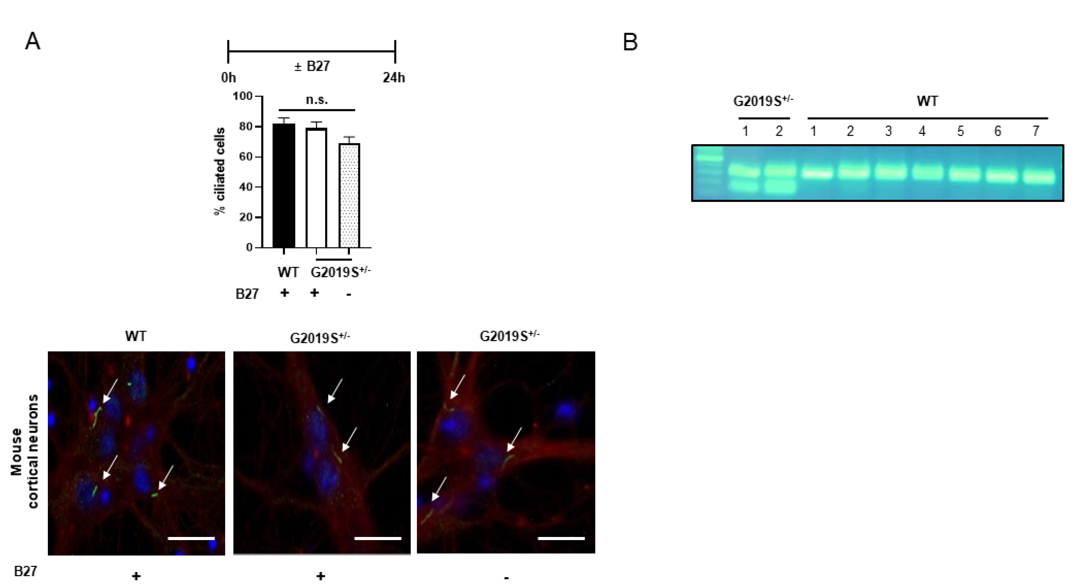
Fig. 2. Ciliogenesis of primary neurons derived from LRRK2 G2019S mice. Murine primary cortical neurons were prepared from LRRK2 G2019S+/- mice and WT littermates. Neurons were exposed to B27 deprivation to mimic serum starvation for 24 h on Day 9. Ciliated neurons were confirmed by positive immunostaining with Arl13B (green) and βIII tubulin (red). (A) The treatment scheme, summary graph, and a representative image for each cell type are shown. A total of 64~100 cells in 8~11 images were analyzed. Scale bar: 20 μm. The white arrow indicates a ciliated cell. (B) Genotyping of each littermate by PCR of tail DNA.
© Exp Neurobiol