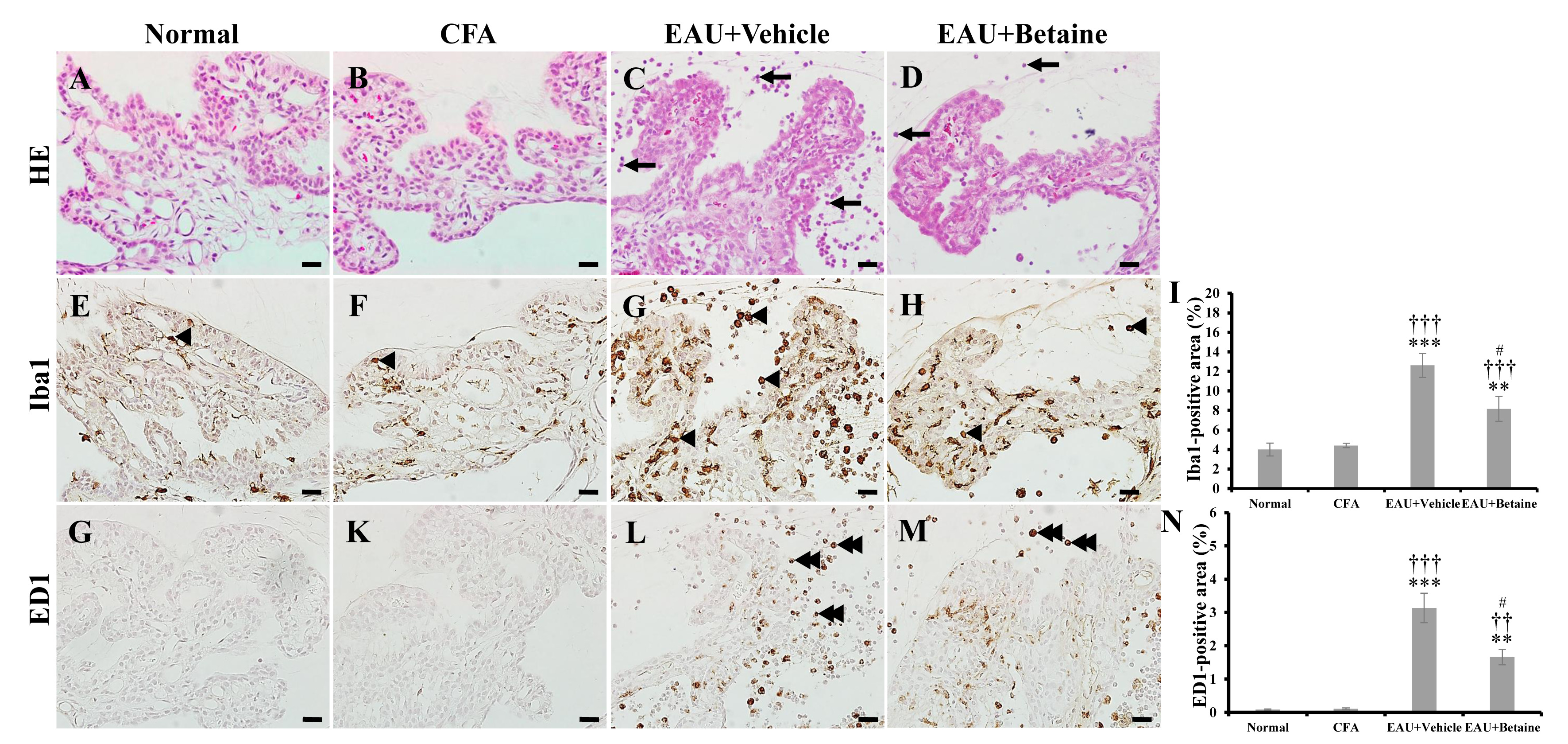
Fig. 3. Histopathological examination of ciliary bodies in the normal (A, E, G), CFA (B, F, K), EAU+Vehicle (C, G, L), and EAU+Betaine (D, H, M) groups. Some rounded cells (arrows in C, D) had infiltrated around the ciliary body in an EAU-induced rat (C, D) but not in normal (A) or CFA (B) rats. An ionized calcium-binding adapter molecule 1 (Iba1)-positive immunoreaction was observed in the ciliary body (arrowheads in E, F, G, H). (I) A significant increase in the Iba1-positive area was confirmed using semi-quantitative analysis. Although the ED1-positive immunoreaction was localized at the infiltrated round cells (double arrowheads in L, M) in the EAU-induced rats, the ciliary bodies of rats in the normal and CFA groups had no ED1-positive cells. (N) The ED1-positive area decreased in response to betaine treatment. Scale bars, 50 μm. **p<0.01; ***p<0.001 vs. normal control; ††p<0.01; †††p<0.001 vs. CFA; #p<0.05 vs. EAU+Vehicle.
© Exp Neurobiol