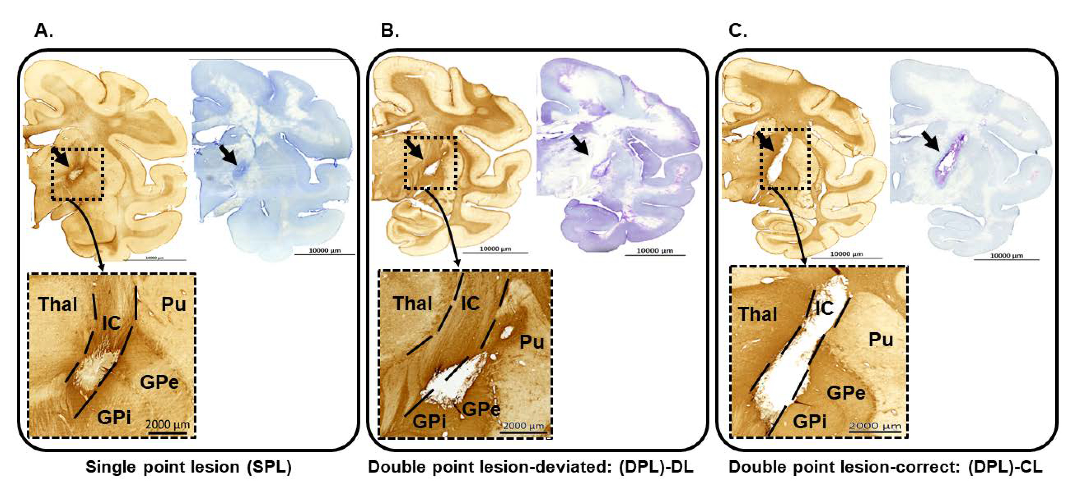
Fig. 4. Histological sections showing the extent of the infarct lesion in each group. GFAP (left) and nissle (right) staining expressed an infarct lesion in the ipsi-lesional hemisphere (×200, top), and the enlarged images were shown at the bottom. Scale bars, 10,000 um (top), 2,000 um (bottom). (A) Single point lesion. (B) Double point lesion – deviated lesion group. (C) Double point lesion – correct lesion group. Thal, thalamus; IC, internal capsule, Pu, putamen; GPe, globus pallidus externus; GPi, globus pallidus internus.
© Exp Neurobiol