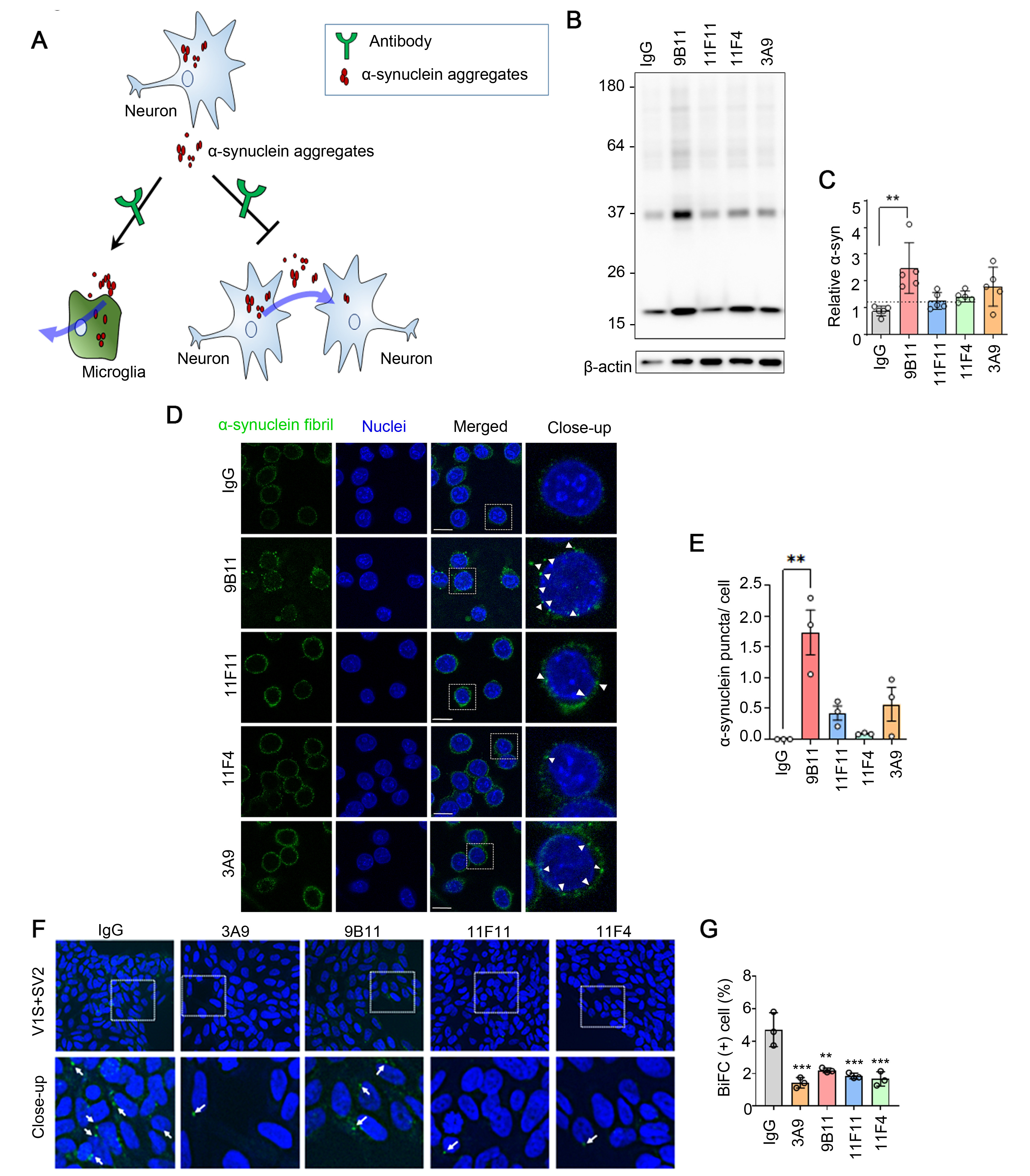
Fig. 2. Conformation-specific antibodies enhance the clearance of extracellular α-synuclein aggregates in BV-2 cells and block cell-to-cell propagation. (A) Proposed mechanism of action of antibodies in the uptake of extracellular α-synuclein and cell-to-cell propagation. (B) Internalization of α-synuclein fibrils in the presence of the indicated antibodies in BV-2 microglial cells. (C) Amount of internalized α-synuclein, quantified and normalized to the levels of β-actin. (D) Immnofluorescence images of α-synuclein fibrils in BV-2 cell after acid wash. Nuclei was stained by DAPI. The close-up panel: Magnified images of the boxed area. Scale bars: 20 μm. (E) Fluorescence puncta from the internalized α-synuclein fibril in (D) were quantified. Puncta was indicated with white arrowheads. One hundred cells per each coverslip were analyzed from three independent experiments. (F) Blocking effects of antibodies against the propagation of α-synuclein. BiFC-positive inclusions are indicated with white arrowheads. Bottom panels: Magnified images of the boxed area. Images were acquired with an InCell analyzer 2200. (G) Quantification of BiFC-positive cells in (F). All data are expressed as means±s.e.m. (n>10,000 cells per experiment (three independent experiments were performed); one-way ANOVA with two-sided Tukey’s post hoc test).
© Exp Neurobiol