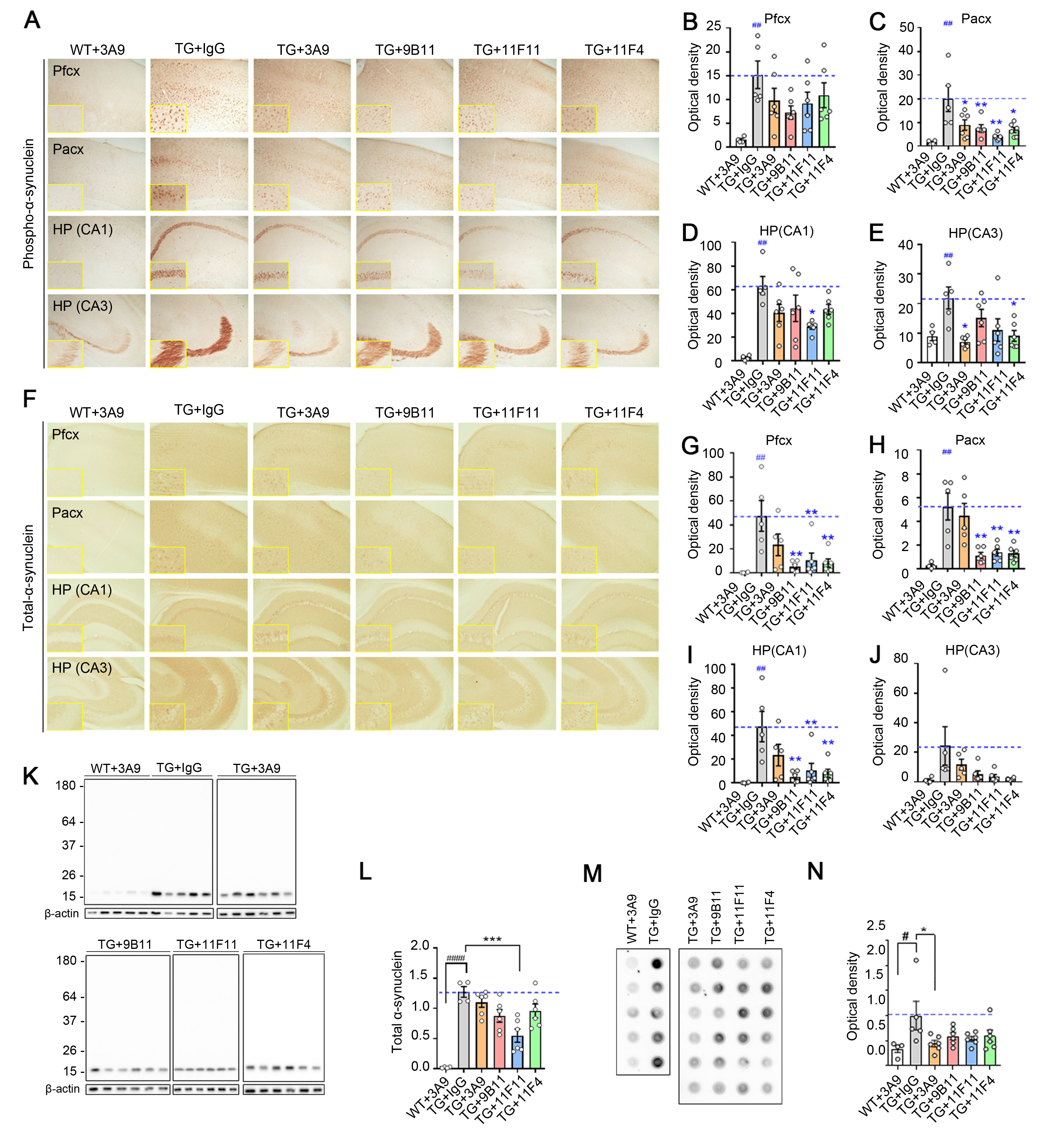
Fig. 3. Passive immunization reduces α-synuclein deposition in mThy-1-α-synuclein Tg mice. mThy-1-α-syn Tg mice and non-Tg littermates were injected with aggregate-specific antibodies and a pan-α-synuclein antibody (10 mg kg-1, i.p.) weekly for 3 months. (A) Representative images of phosphorylated α-synuclein (pS129) staining in the prefrontal cortex, parietal cortex, and hippocampal regions. (B~E) Optical density measurements in the prefrontal cortex (B), parietal cortex (C), and hippocampal regions (D, E). (F) Representative images of total-α-synuclein staining in the prefrontal cortex, parietal cortex, and hippocampal regions. (G~J) Optical density measurements in the prefrontal cortex (G), parietal cortex (H), and hippocampal regions (I, J). Scale bar, 200 μm, Data are expressed as means±s.e.m. (n=5~6 per group; #p<0.05 vs. WT+3A9 group, *p<0.05 vs. TG+IgG group; one-way ANOVA with two-sided Tukey’s post hoc test). Pfcx, prefrontal cortex; Pacx, parietal cortex; HP, hippocampus. (K~L) Western blotting of brain tissue extracts. (M~N) Dot blotting of brain tissue extracts. FILA-4 antibody, an antibody specific for fibrillary α-synuclein were used. Data were analyzed by one-way ANOVA with two-sided Tukey’s post hoc test. *p<0.05 ****p<0.0001 vs. TG+IgG group.
© Exp Neurobiol