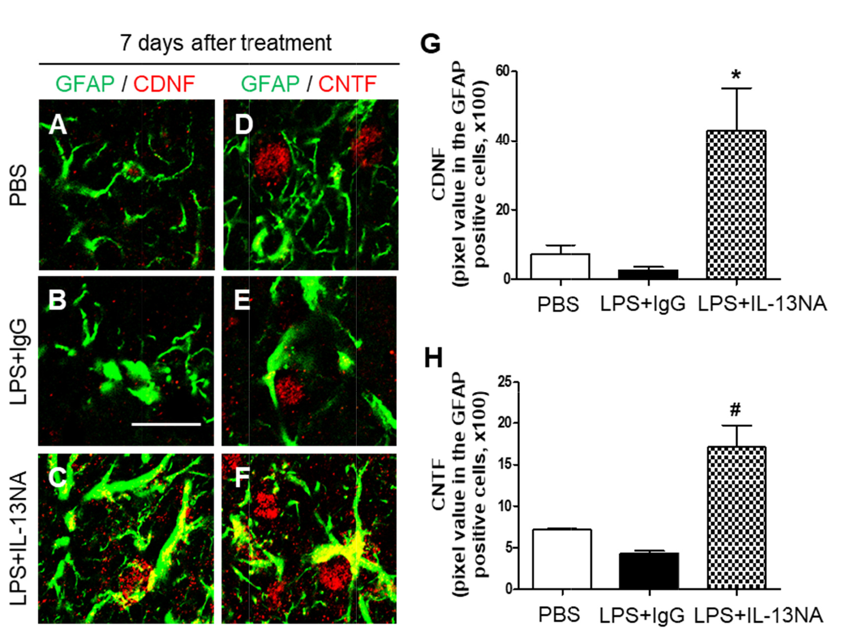
Fig. 6. Interleukin-13 inhibits expression of neurotrophic factors on astrocytes in LPS-injected rat striatum
in vivo. Sections (A, D, PBS; B, E, LPS+IgG; C, F, LPS+IL-13NA) adjacent to those used in
Figure 3 were immunostained with GFAP (A~F) antibody for astrocytes, and cerebral dopamine neurotrophic factor (CDNF: A~C) or ciliary neurotrophic factor (CNTF: D~F) antibodies. (A~C) Immunofluorescence images of GFAP (green) and CDNF (red), and both images are merged (Yellow) in the striatum at 7 days after LPS+IgG injection. (D~F) Immunofluorescence images of GFAP (green) and CNTF (red), and both images are merged (Yellow) in the striatum at 7 days after LPS+IgG injection. (G) Quantification of CDNF expression in GFAP
+ cells in the striatum at 7 days after LPS injection. *p<0.05, as compared with LPS+IgG. One-way ANOVA and Newman–Keuls analyses. Three animals were used for each experimental group. The results represent mean±SEM. (H) Quantification of CNTF expression in GFAP
+ cells in the striatum at 7 days after LPS+IgG injection.
#p<0.01, as compared with LPS+IgG. One-way ANOVA and Newman–Keuls analyses. Three animals were used for each experimental group. The results represent mean±SEM. Scale bar, (A~F) 20 µm.