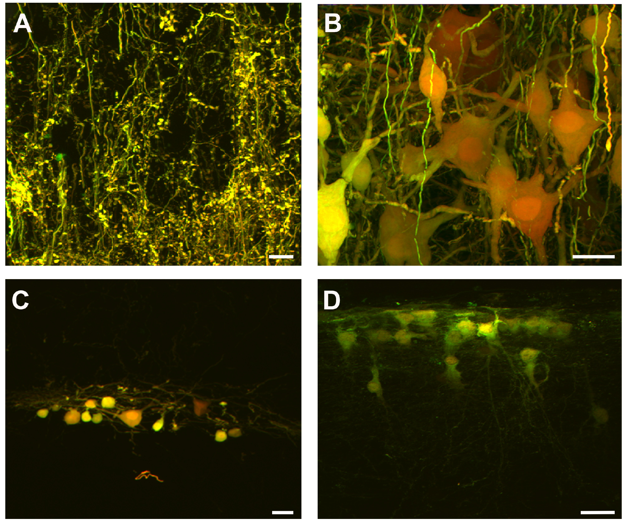
Fig. 3. Images of the spinal cord in L4-L5 segments with the mixture of RDA and FDA fluorescent markers. (A) Terminal afferent fibers arriving at the ventral spinal cord were stained by applying the fluorescent mixture in the L5 dorsal root (scale bar, 10 µm). (B) The mixture was administered through the L5 ventral and dorsal root. It is important to note that the motoneurons and the afferent fiber arriving at the motor nucleus were stained (scale bar, 50 µm). (C) Neurons traveling grouped on the spinal cord dorsal surface. (D) The neurons were penetrating a deep region in the spinal cord.
© Exp Neurobiol