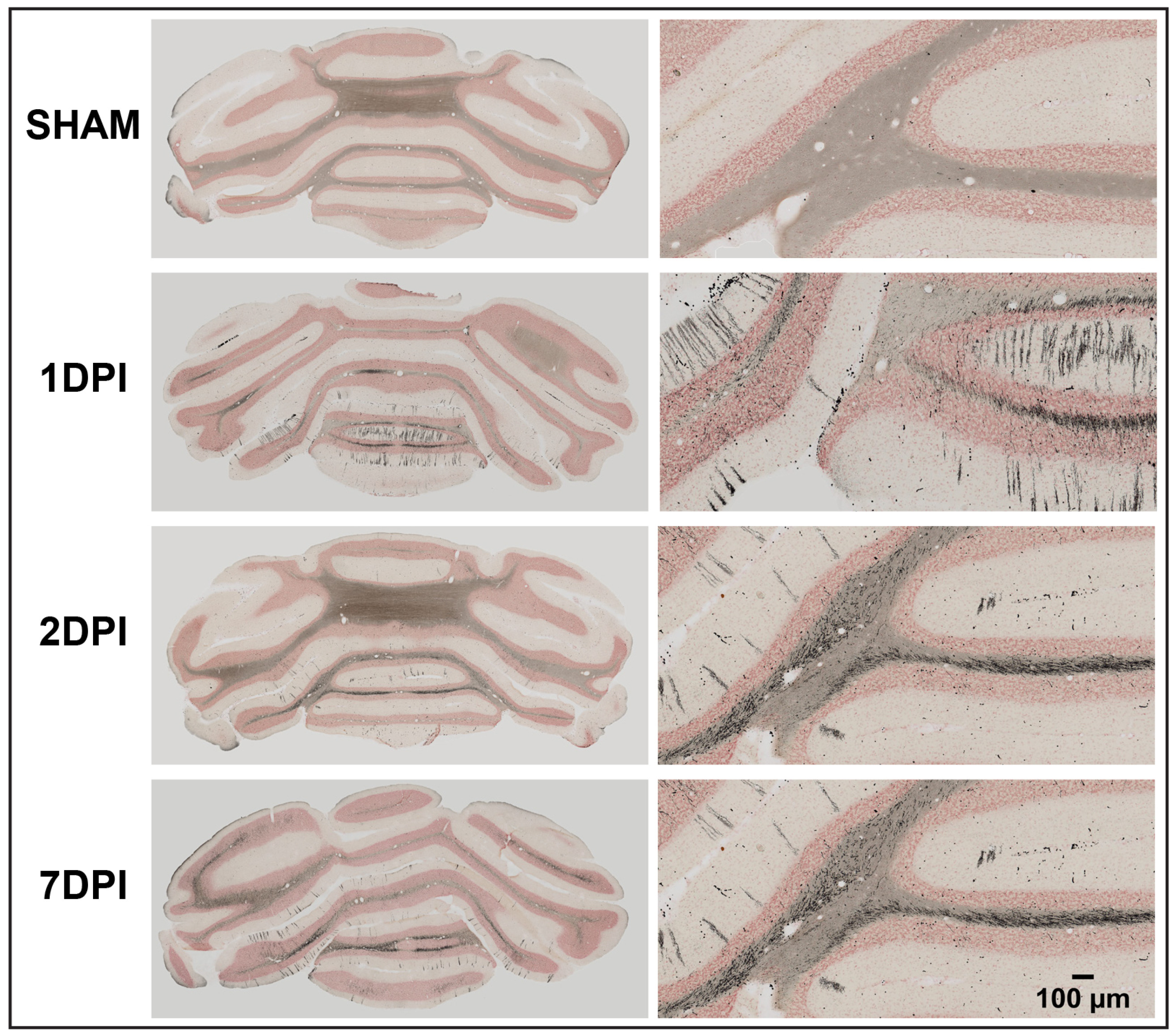
Fig. 2. The cerebellum was montaged for analysis of neuropathology. High magnification representative images show stripes of neuropathology visible in the molecular layer of the cerebellar cortex following diffuse TBI that are not present in sham animals. Additional neuropathology is visible in cerebellar white matter tracts. Images from coronal sections were montaged to include full coronal cerebellar sections in the analysis. Scale bar 100 µm for representative images.
© Exp Neurobiol