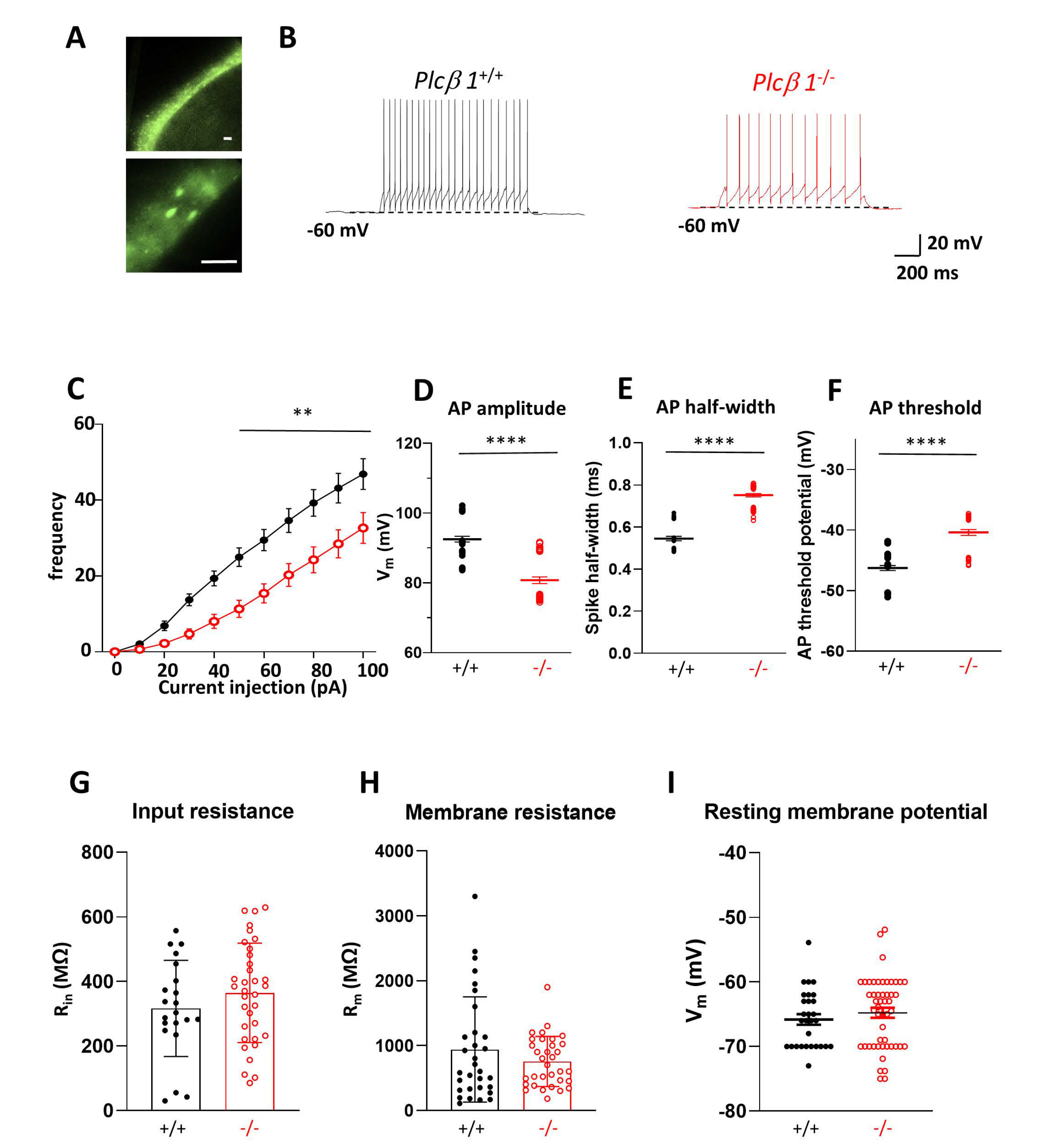
Fig. 4. Tonic firing of the vmTRN is decreased in Plcβ1-/- mice. (A) Recording of vmTRN neurons from Gad65-GFPtg; Plcβ1+/+ or Gad65-GFPtg; Plcβ1-/- mice. (B) Representative traces of tonic firings in Plcβ1+/+ (left) and Plcβ1-/- (right) vmTRN neurons. (C) Average number of tonic firings with different current pulses in Plcβ1+/+ and Plcβ1-/- vmTRN neurons. (D) AP peak height: 92.5±2.0 and 80.8±2.0 mV in Plcβ1+/+ and Plcβ1-/- mice, two-tailed, ****p<0.0001. (E) AP 1/2 width: 0.55±0.2 and 0.75±0.2 ms in Plcβ1+/+ and Plcβ1-/- mice, two-tailed, ****p<0.0001. (F) AP threshold: -46.3±0.6 and -40.4±0.6 mV in Plcβ1+/+ and Plcβ1-/- mice, two-tailed, ****p<0.0001. (G) Input resistance: 316.63±42 and 364.7±42 MΩ in Plcβ1+/+ and Plcβ1-/-, two-tailed, n.s. p=0.25. (H) Membrane resistance: 860.4±136 and 718±136 MΩ in Plcβ1+/+ and Plcβ1-/- mice, two-tailed, n.s. p=0.30. (I) Resting membrane potential: -65.8±1.1 and 64.8±1.1 mV in Plcβ1+/+ and Plcβ1-/- mice, two-tailed, n.s. p=0.38. All data are presented as the mean±SEM.
© Exp Neurobiol