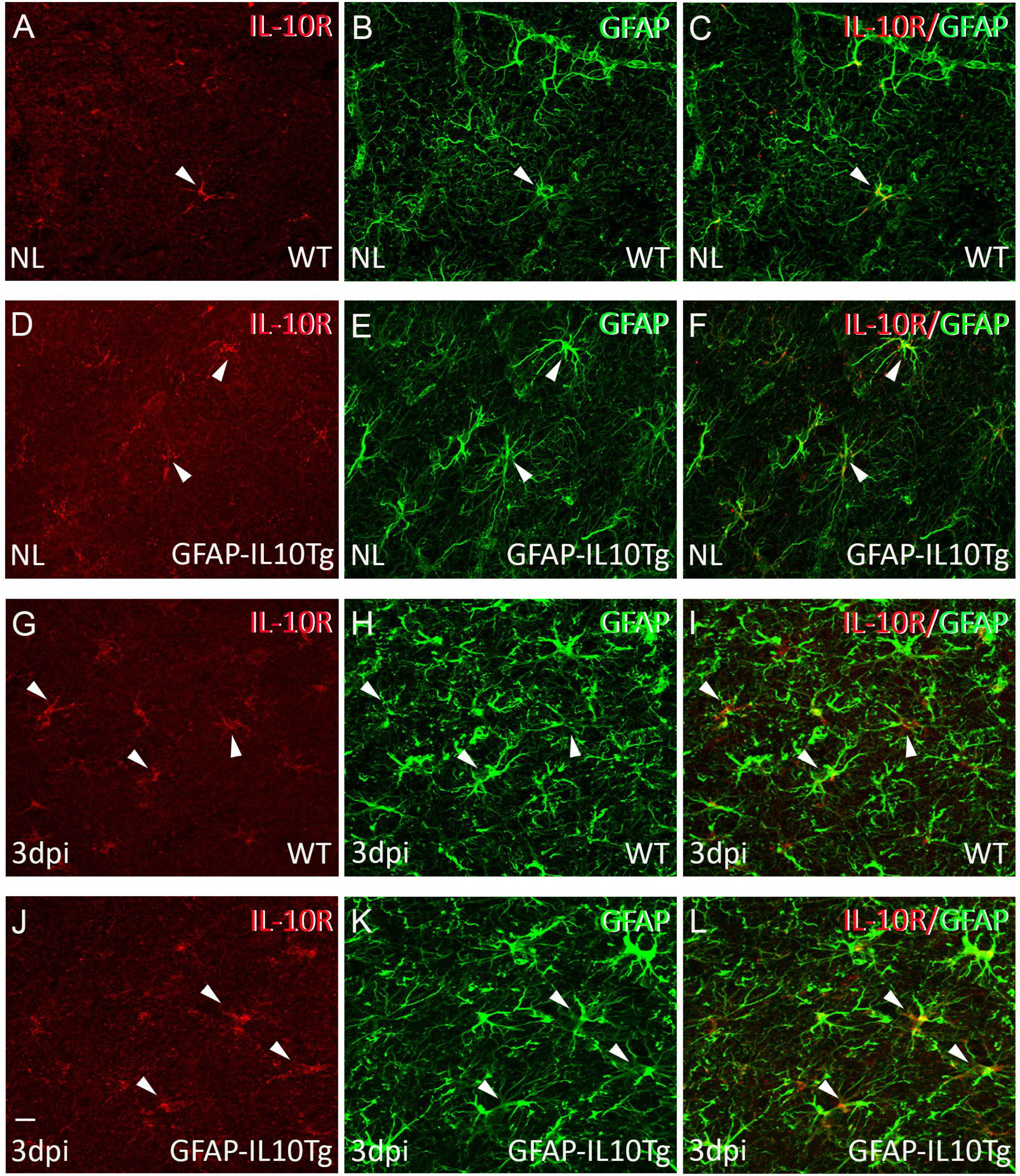
Fig. 12. IL-10R expression. Representative images showing the double immunofluorescence combining IL-10R (red) and GFAP (green) in non-lesioned (NL) (A~F) and TBI-lesioned animals at 3 dpi (G~L). Note that a major part of IL-10R+ cells showed co-localization (yellow) with GFAP in both NL and after TBI in both genotypes (arrowheads). Scale bar=10 µm.
© Exp Neurobiol