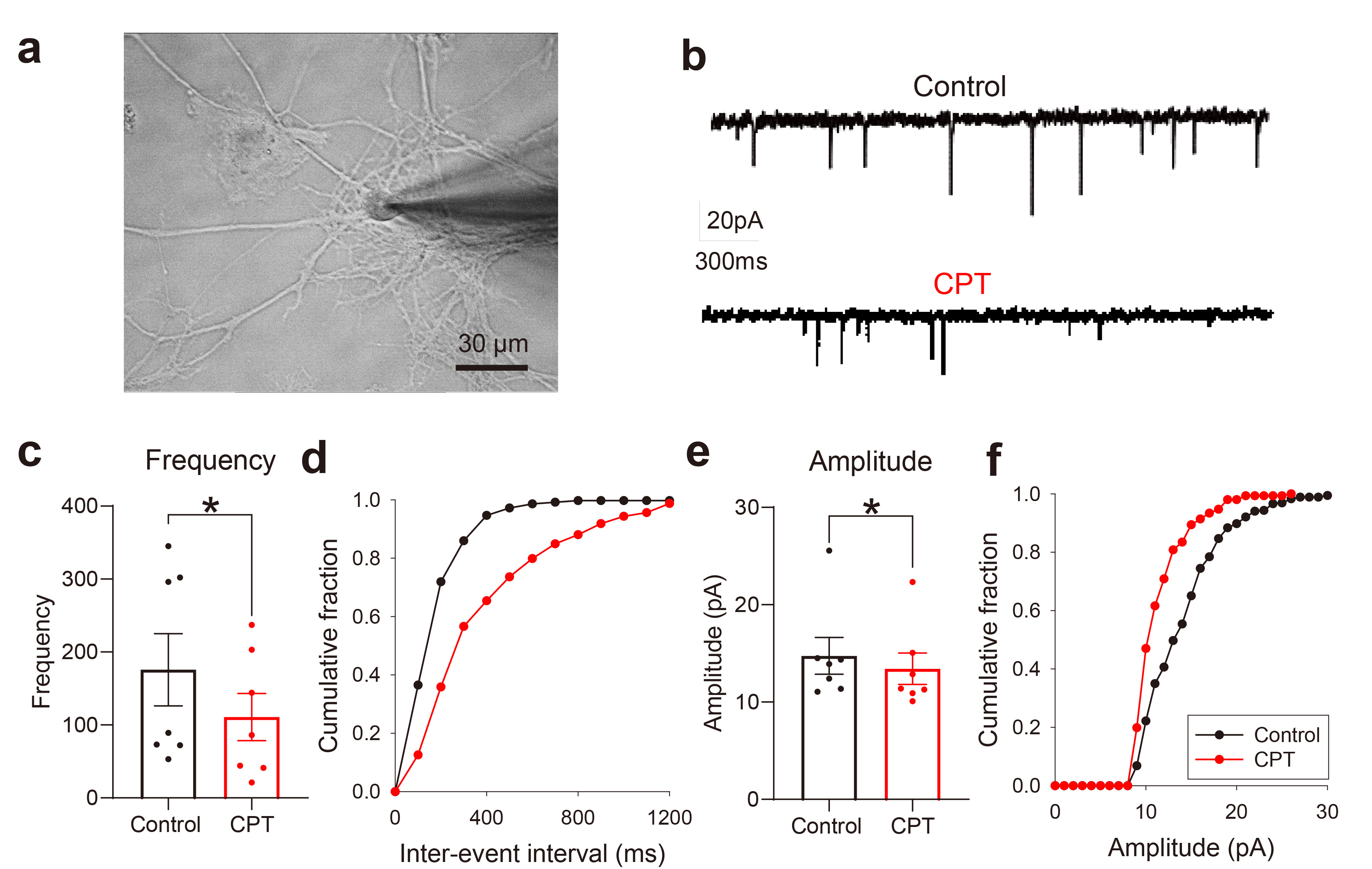
Fig. 1. CPT reduces both the frequency and amplitude of sEPSCs in rat hippocampal neurons. (a) Bright field image showing a glass pipette used for whole-cell patch-clamp recording of primary cultured rat hippocampal neurons. (b) Representative sEPSCs before (upper panel) and during the treatment with 10 µM CPT (lower panel). (c) Bar graph summarizing the frequencies (paired t test, *p=0.04, n=7). (d) Cumulative fraction of the interevent interval before (black line) and during the treatment with 10 µM CPT (red line). (e) Bar graph summarizing the amplitudes (paired t test, *p=0.04, n=7). (d) Cumulative fraction of the amplitude before (black line) and during the treatment with CPT (red line).
© Exp Neurobiol