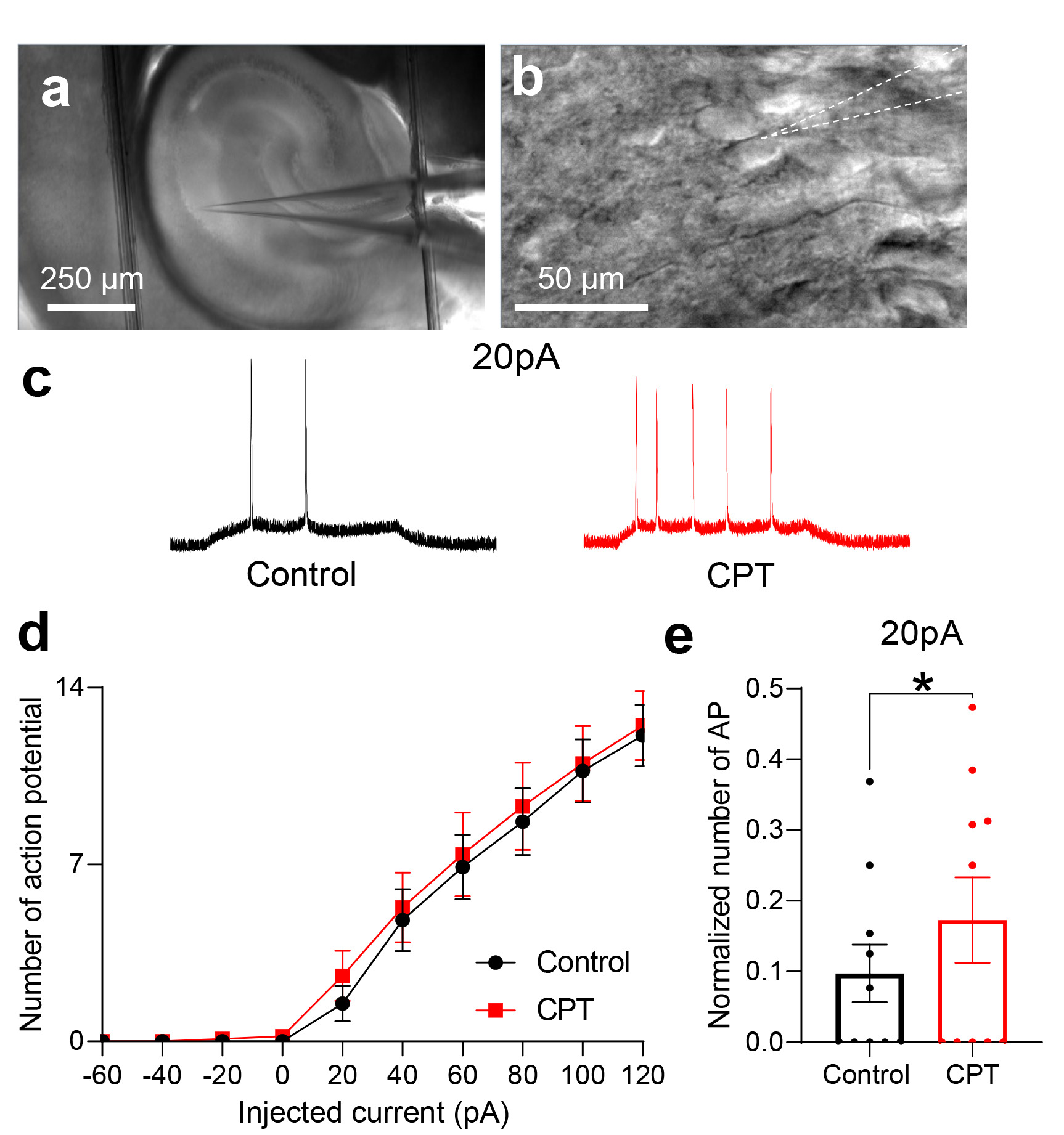
Fig. 2. CPT increases the number of APs generated by application of a +20-pA current to hippocampal CA1 pyramidal neurons from C57BL/6 mice. (a) Bright field image showing a glass pipette above the hippocampal CA1 region. (b) High-magnification image showing a glass pipette used for the whole-cell patch-clamp recording of CA1 pyramidal neurons. (c) Representative voltage trace in the current clamp mode before the treatment (left panel, black trace) and during the treatment with 10 µM CPT (right panel, red trace). (d) Plot comparing the numbers of APs recorded during a 0.6 s current application using the current step protocol (black indicates before the CPT treatment; red indicates during the CPT treatment). (e) Summary bar graph comparing the numbers of APs recorded before and during the treatment with CPT (paired t test, *p=0.04, n=10).
© Exp Neurobiol