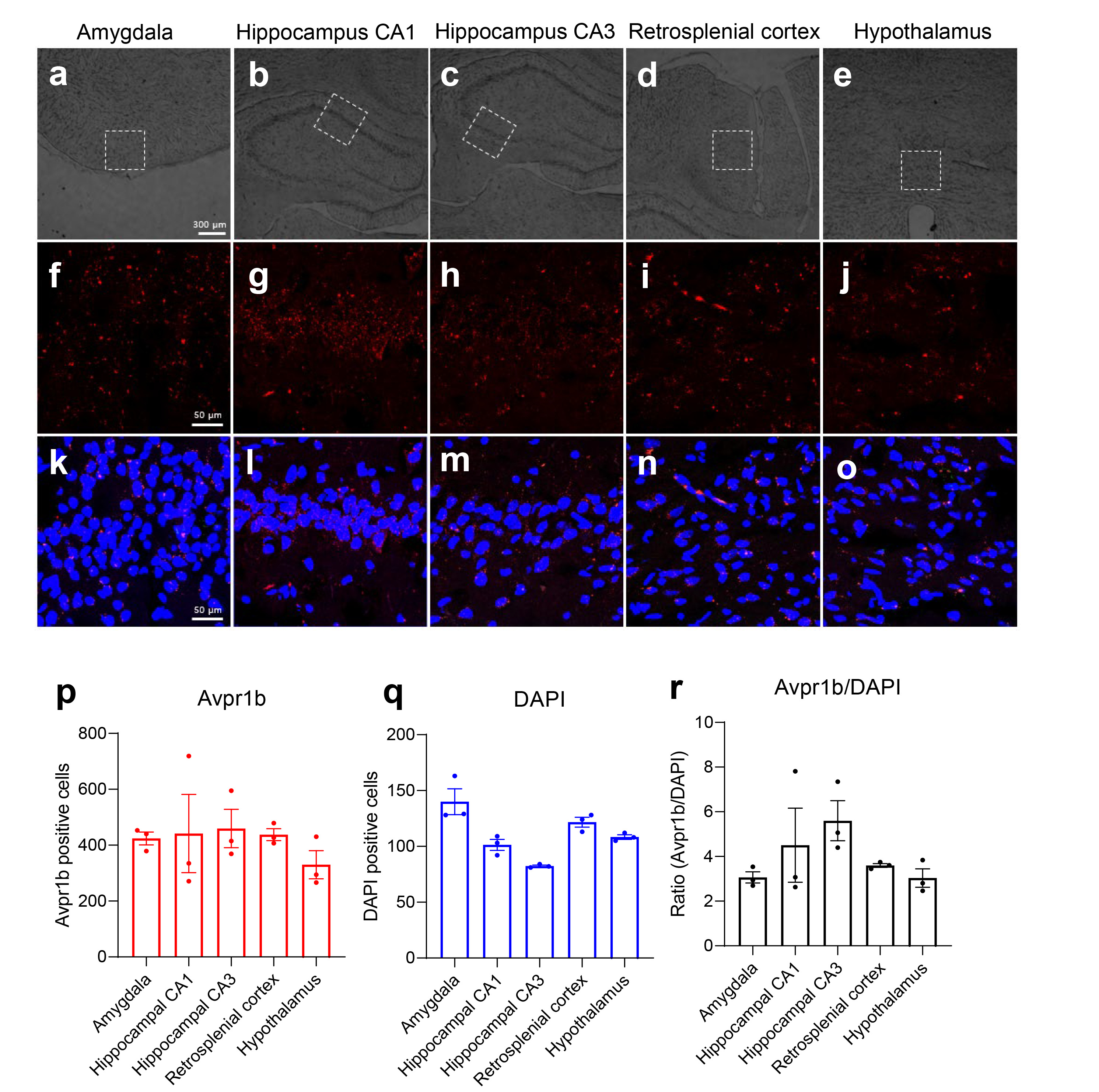
Fig. 5. Avpr1b was generally expressed at high levels in the hippocampus. (a) Bright field images of the amygdala, (b) hippocampal CA1 region, (c) hippocampal CA3 region, (d) retrosplenial cortex, (e) hypothalamus. (f) Avpr1b expression in the amygdala. (g) Avpr1b expression in the hippocampal CA1 region. (h) Avpr1b expression in the hippocampal CA3 region. (i) Avpr1b expression in the retrosplenial cortex. (j) Avpr1b expression in the hypothalamus. (k) DAPI and Avpr1b staining in the amygdala. (l) DAPI and Avpr1b staining in the hippocampal CA1 region. (m) DAPI and Avpr1b staining in the hippocampal CA3 region. (n) DAPI and Avpr1b staining in the retrosplenial cortex. (o) DAPI and Avpr1b staining in the hypothalamus. (p) Bar graph summarizing the number of Avpr1b puncta. (q) Bar graph summarizing the number of DAPI-stained. (r) Ratio (number of red puncta/number of blue-stained regions) showing that Avpr1b was expressed at high levels in the hippocampus.
© Exp Neurobiol