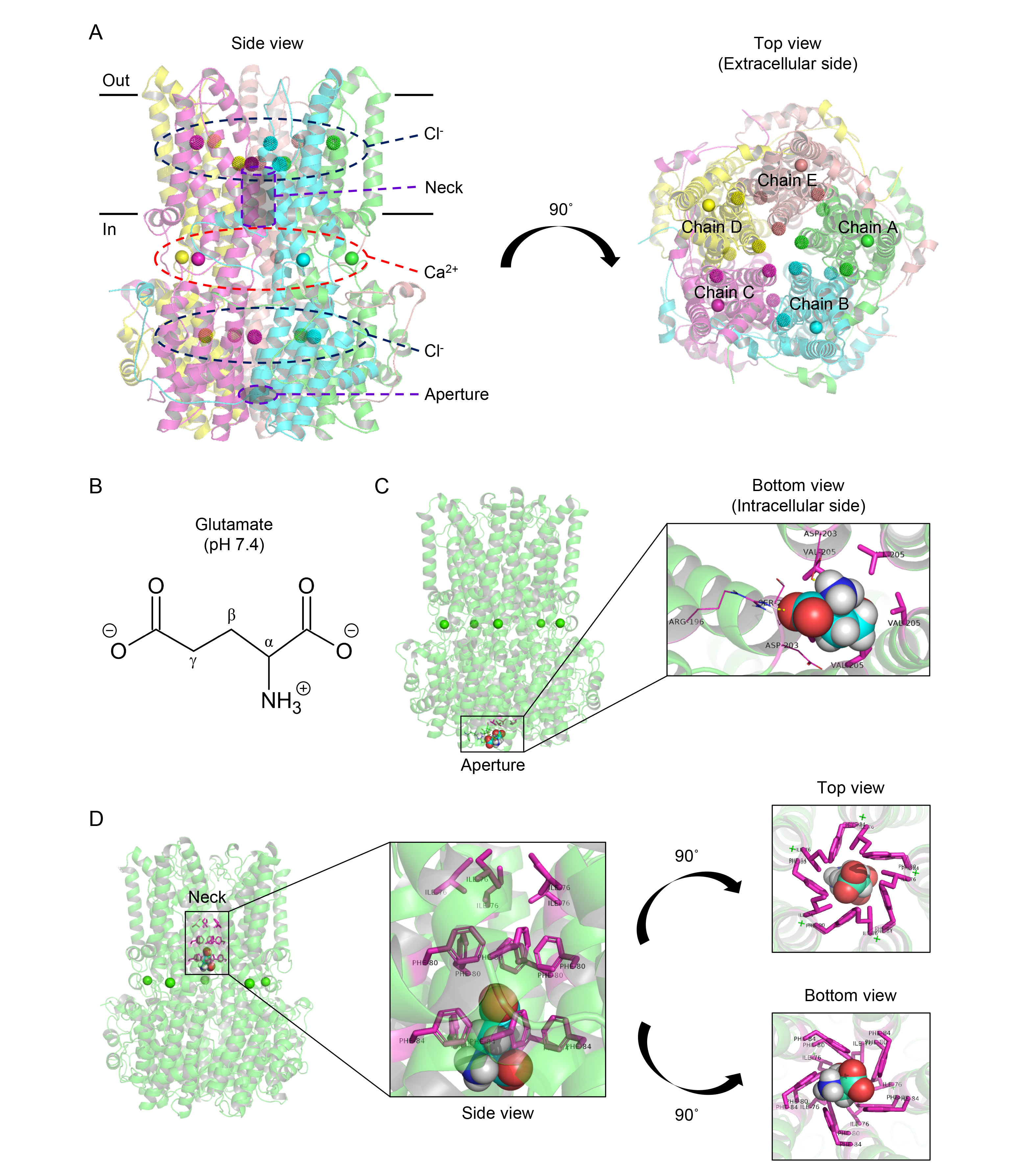
Fig. 1. Glutamate is docked on the aperture and neck of the cBest1. (A) Side view (left) and top view (right) showing modified X-ray crystal structure of pentameric cBest1 with Cl- and Ca2+ (amino acids 1-405; PDB ID code: 4RDQ). (B) 2D structure of glutamate at pH 7.4. (C) Docked pose of glutamate at the aperture. The cBest1 is depicted as a Ca2+-bound (green sphere) structure (transparent green ribbon diagram). Glutamate and V205 residue are represented by the sphere model (carbon, cyan; oxygen, red; nitrogen, blue; hydrogen, white) and the stick model (magenta), respectively. (D) Docked pose of glutamate at the neck. I76, F80, and F84 residues are represented by the stick model.
© Exp Neurobiol