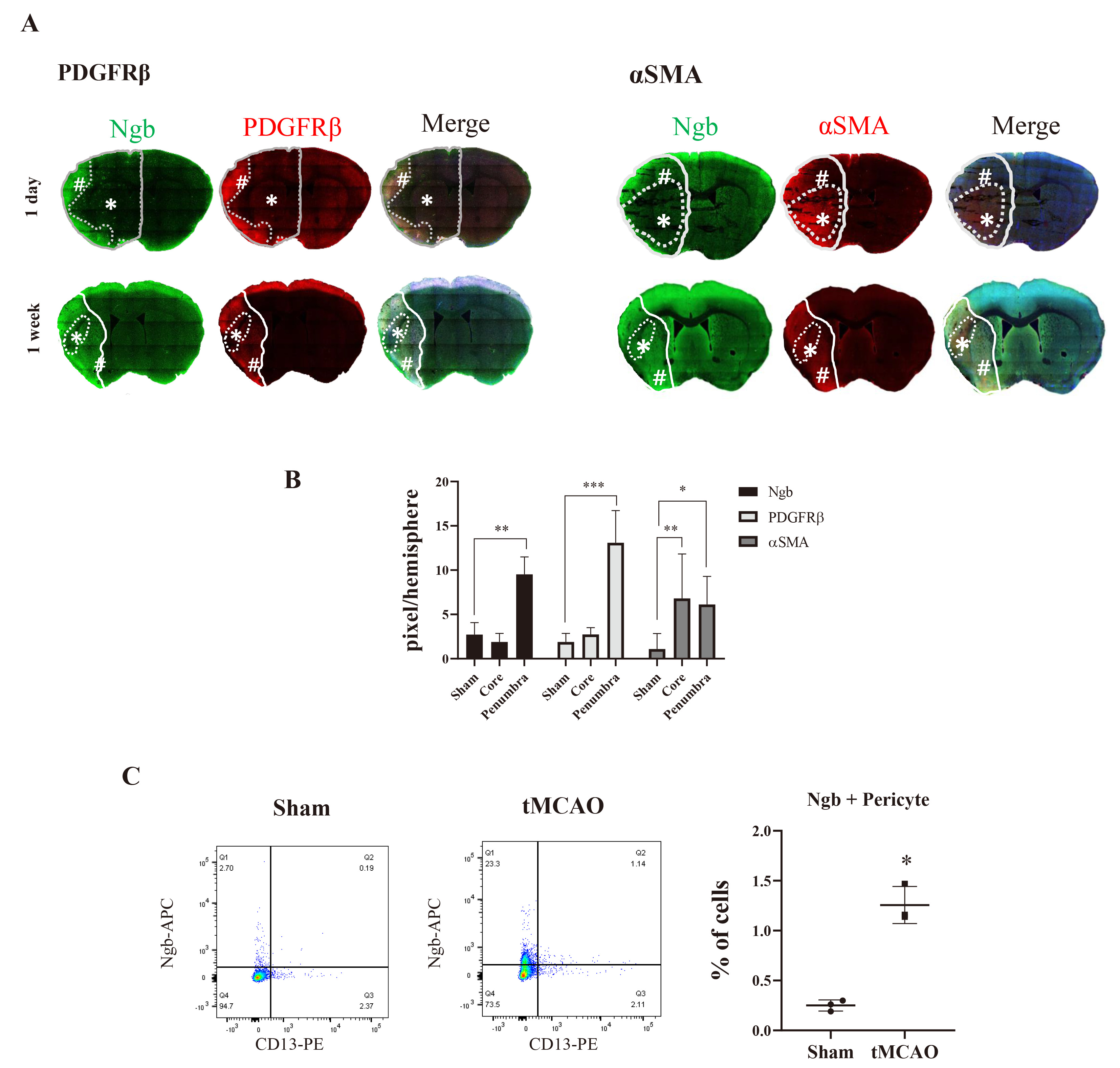
Fig. 3. Different distribution of Ngb in different subtypes of pericytes after stroke. (A) The increased level of Ngb was colocalized with PDGFRβ-positive pericytes in the penumbra of the infarct area 1 day and 1 week after tMCAO. However, it was decreased in the core region. In contrast, αSMA-positive pericyte level was high in the core, which was not colocalized with that of Ngb after tMCAO at 1 day and 1 week (*: core, #: penumbra). (B) Quantification of tile scan image of 1 day. Values are expressed as mean±SD (n=4) (*p<0.05, **p<0.01, ***p<0.001, relative to sham by two-way ANOVA). (C) The amount of pericyte expressing Ngb in the whole brain was confirmed by FACS after tMCAO in mice. The pericytes expressing Ngb were increased in the ipsilateral side after tMCAO. Ngb-expressing cells increased by 5-fold from the ipsilateral side, compared with the sham group. Quantification of Ngb-positive pericytes. (mean±SD, n=3) *p<0.05, relative to sham by t-test.
© Exp Neurobiol