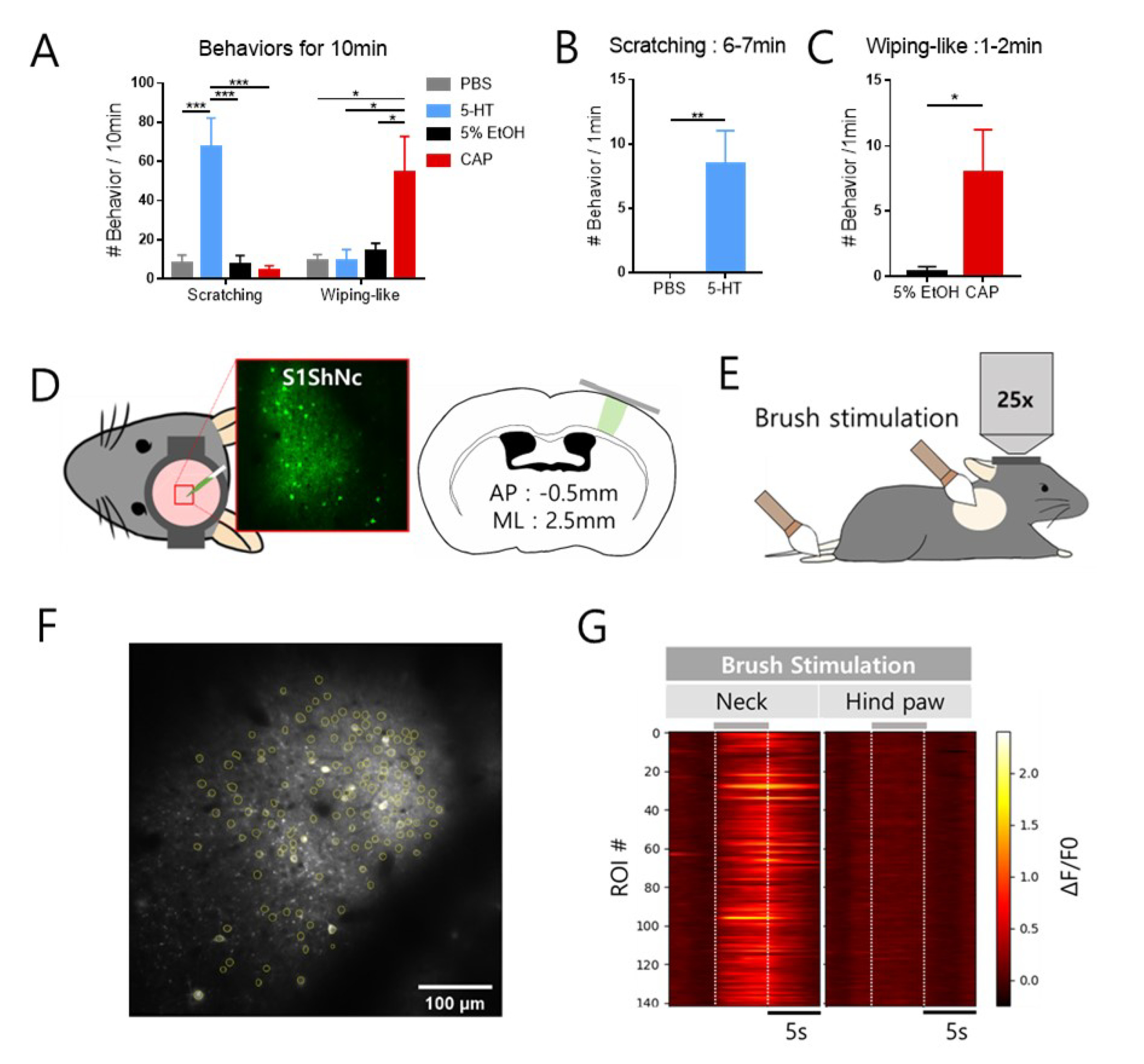
Fig. 1. In vivo two-photon calcium imaging of S1ShNc neurons. (A) Behavioral observation for itch and pain were performed in mice for 10 min. One-way ANOVA test was performed with Tukey’s post hoc for multiple comparisons. (B) Scratching behaviors evoked by intradermal injection of PBS (n=7) or 5-HT (n=8) on the lateral side of neck for 6~7 min after treatment (unpaired t-test). (C) Wiping-like behaviors evoked by intradermal injection of 5% EtOH (n=8) or CAP (n=7) on the lateral side of neck for 1~2 min after treatment (unpaired t-test). (D) Craniotomy were performed over the left S1 cortex, and GCaMP6s virus were expressed in layer 2/3 neurons of S1ShNc region. The S1ShNc region is located laterally 2.5 mm and posteriorly 0.5mm from bregma. Area marked in green of the coronal section image indicate the S1ShNc region. (E) To identify whether the S1ShNc is corresponding region of lateral side of neck, we recoded Ca2+ activities in anesthetized mice during brush stimulation to the right side of neck or hind paw. (F) Representative images of Ca2+ fluorescence of the S1ShNc neurons in response to brush stimulation for left side of neck, yellow lines indicate the ROIs that semi-automatically identified. (G) Representative color-coded raster plots of S1ShNc neurons in response to mechanical neck or hind paw stimulation. Stimulation was applied for 5 s and gray bars over the plot indicate the point of stimulation. Data are expressed as mean SEM (*p<0.05, **p<0.001, and ***p<0.0001).
© Exp Neurobiol