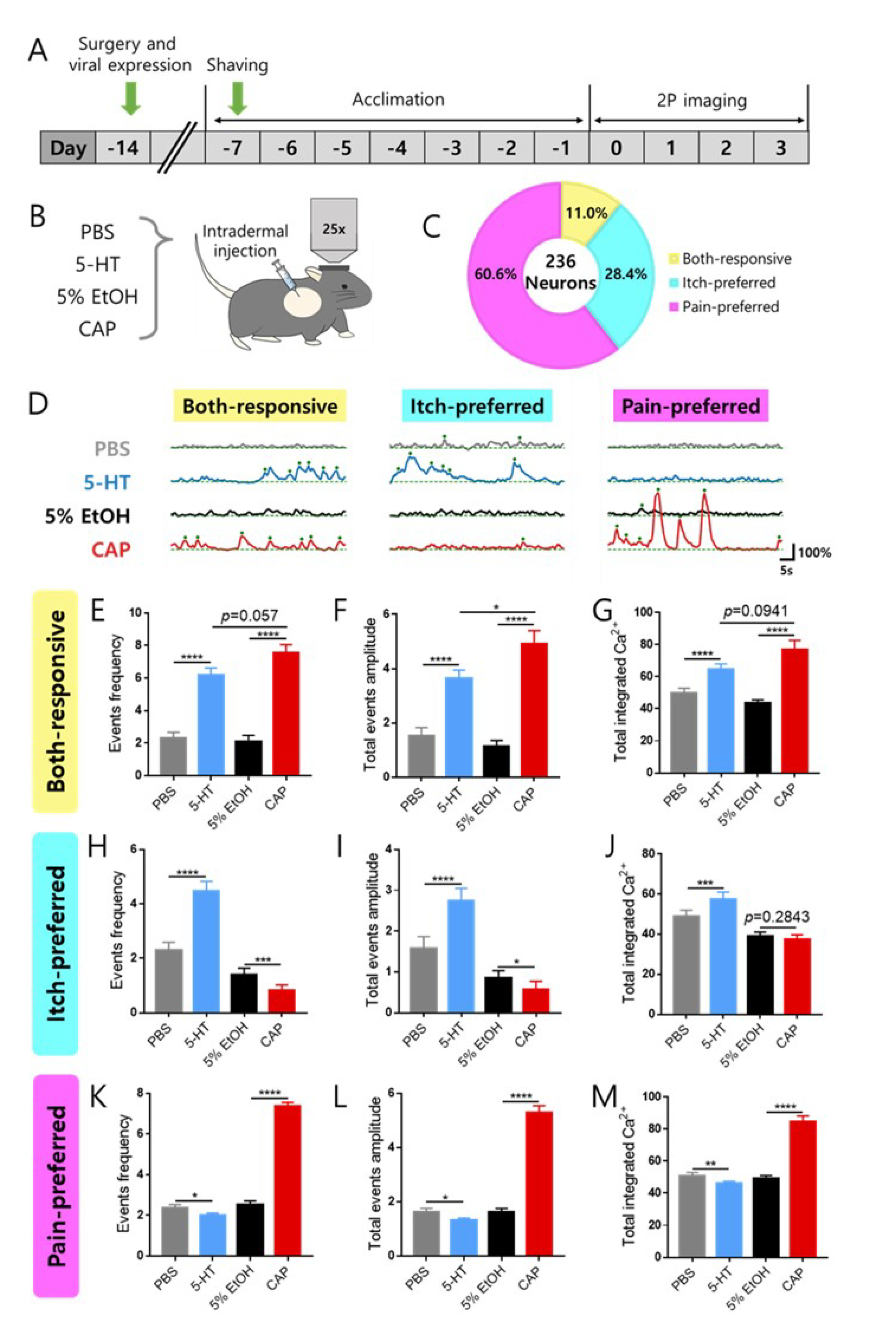
Fig. 2. Classification of itch and pain selective neurons according to changes in spontaneous calcium response. (A, B) Schedule and schematic illustration for two photon imaging for awake head fixed mice. A total of 1271 cells were imaged from eight mice. Among those cells, 236 cells were responsive for itch or condition. (C) Pie chart for responsive neurons for itch or pain conditions. (D) Representative Ca2+ traces for classified cell populations. We marked the Ca2+ traces for four different conditions with distinct colors. Gray, blue, black, and red indicate the activity during the PBS, 5-HT, 5% EtOH, and Cap sessions, respectively. (E~G) Response properties for both-responsive neurons. (H~J) Response properties for itch-preferred neurons. (K~M) Response properties for pain-preferred neurons. Data are expressed as mean SEM (paired t test; *p<0.05, **p<0.01, ***p<0.001, and ****p<0.0001).
© Exp Neurobiol