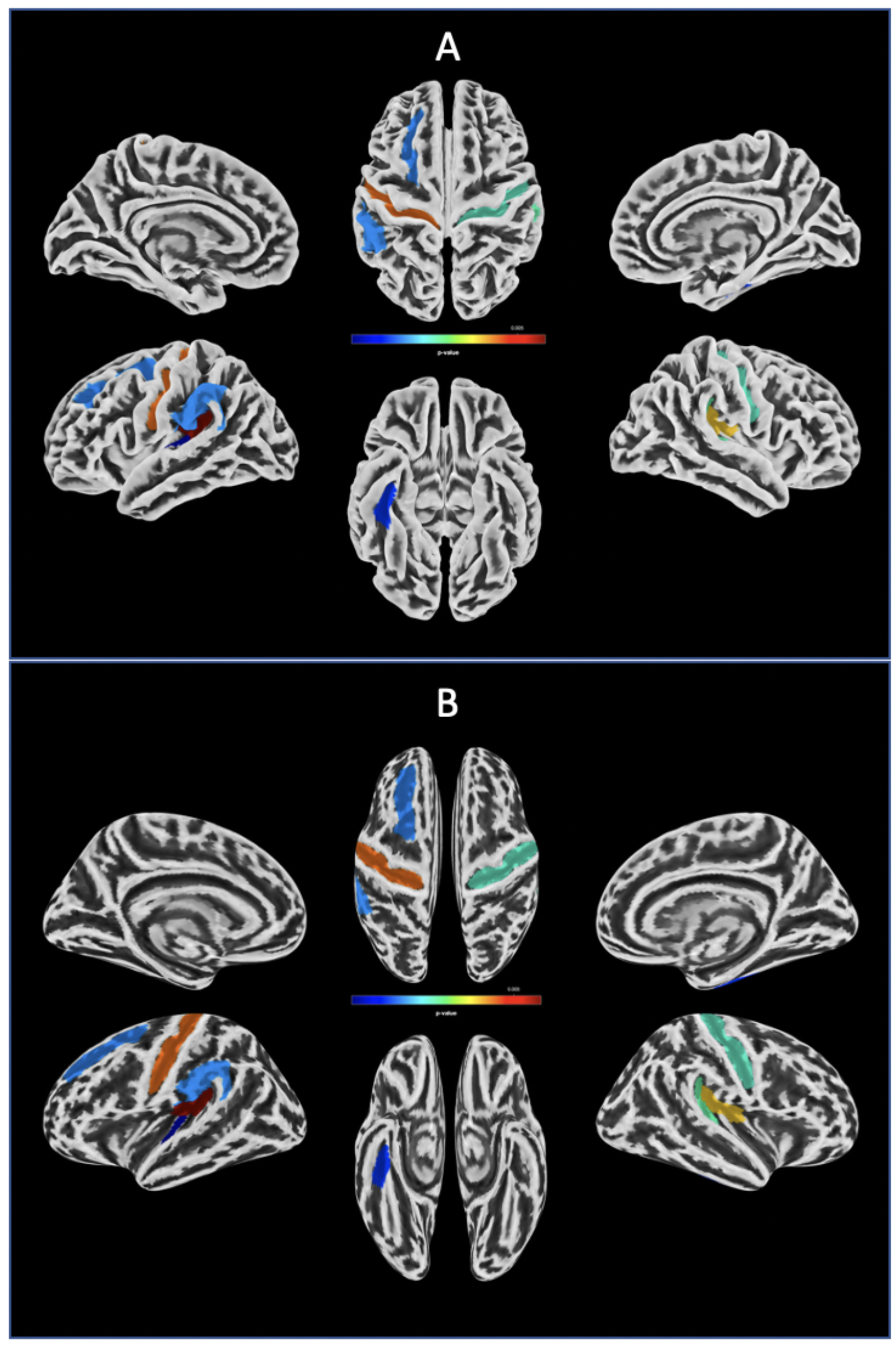
Fig. 1. Reduced sulcal depth in MDD patients. Sulcal depths of MDD were reduced compared to the normal controls in the left hemisphere of posterior ramus of the lateral sulcus (Lat_Fis-post), superior frontal sulcus (S_front_sup), supramarginal gyrus (G_pariet_inf-Supramar), central sulcus (S_central), Heschl's gyrus (G_temp_sup-G_T_transv), and in the right hemisphere of Lat_Fis-post, temporal plane of the superior temporal gyrus (G_temp_sup-Plan_tempo), anterior transverse collateral sulcus (S_collat_transv_ant), and Heschl's gyrus. The statistical result was displayed in aparc a2009s atlas (A) and template brain was inflated to show the areas of sulcal depth in detail (B). The statistical levels were set to a Holm-Bonferroni corrected p<0.05 level.
© Exp Neurobiol