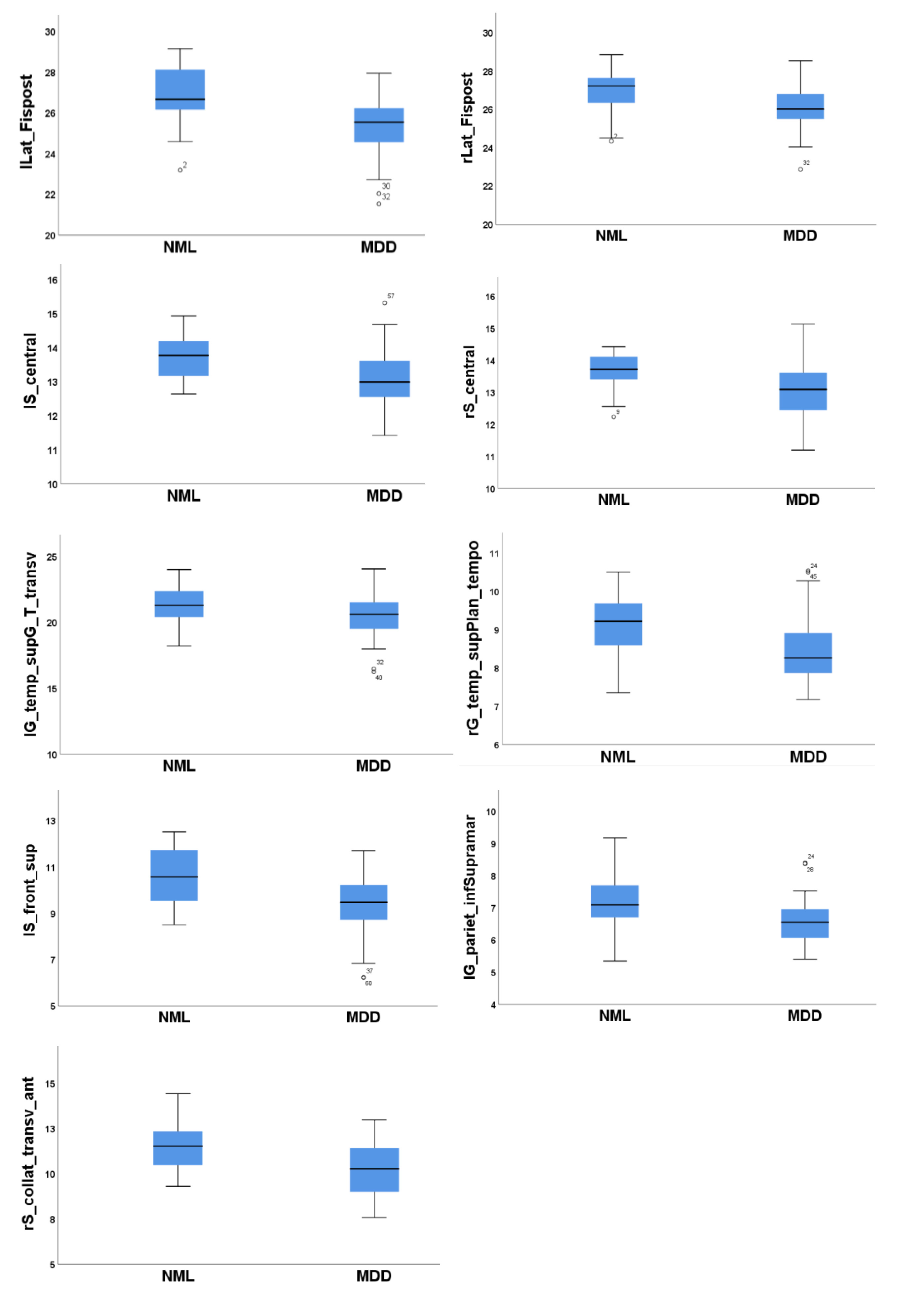
Fig. 2. Box and whisker plots of significantly reduced sulcal depth in depressed patients. Left posterior ramus of the lateral sulcus (lLat_Fispost), right posterior ramus of the lateral sulcus (rLat_Fispost), left central sulcus (lS_central), right central sulcus (rS_central), left Heschl’s gyrus (lG_temp_supG_T_transv), right temporal plane of the superior temporal gyrus(rG_temp_sup_plan_tempo), left superior frontal sulcus (lS_front_sup), left supramarginal gyrus (lG_pariet_inf-Supramar), right anterior transverse collateral sulcus (rS_collat_transv_ant).
© Exp Neurobiol