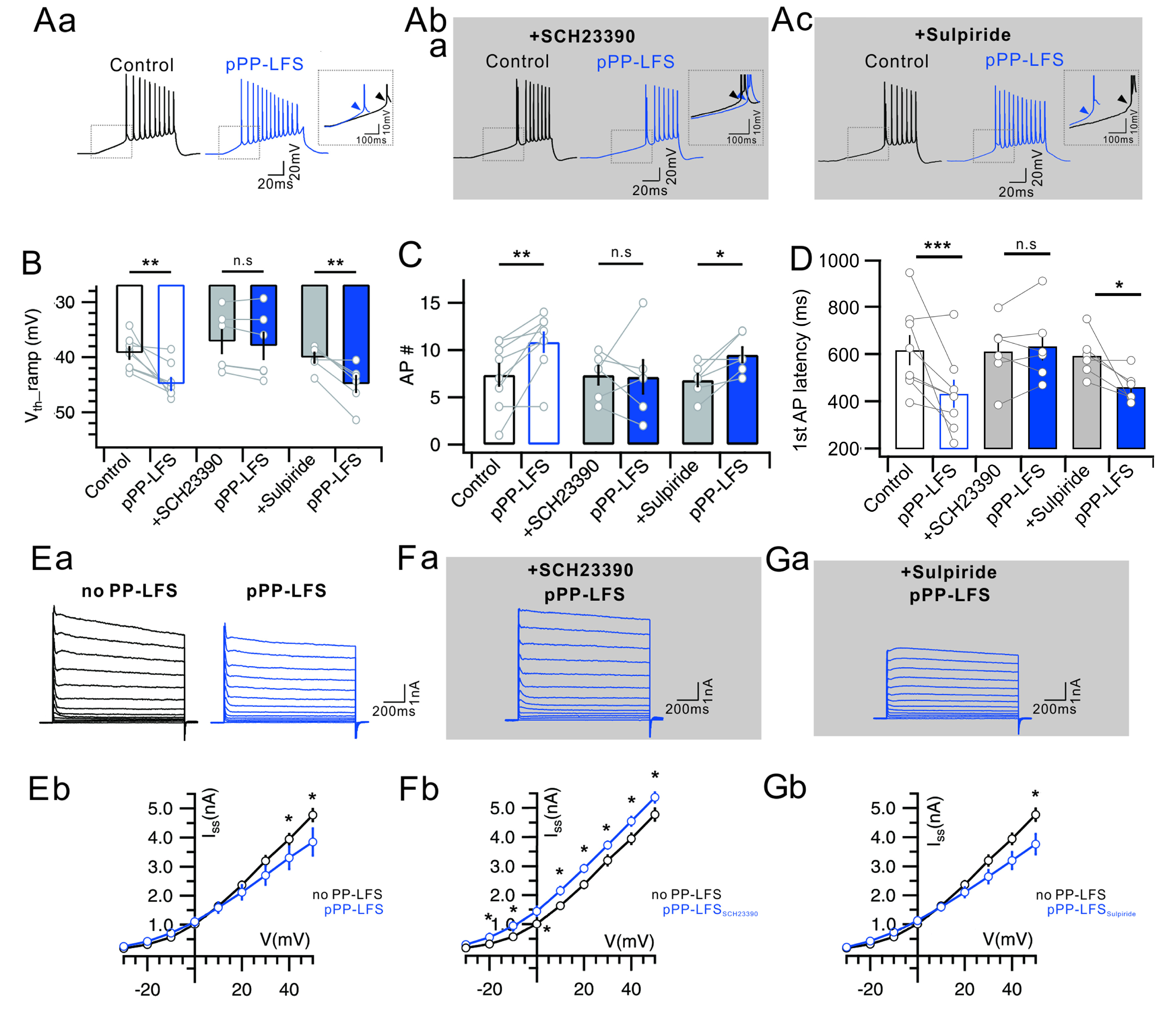
Fig. 3. D1-dependent decrease in the K
+ current underlies the PP-LFS induced the E-S potentiation. (Aa) Representative voltage responses to a ramp (250 pA/s) current injection to soma before (Control) and 30 min after PP-LFS (pPP-LFS).
Inset: Superimposed traces of significant V
th hyperpolarization and shortened latency of the 1
st AP. (Ab) Representative voltage responses to a ramp (250 pA/1 s) current injection to soma for the control and the pPP-LFS during the bath application of SCH23390.
Inset: Superimposed traces of significant V
th hyperpolarization and shortened latency of the 1
st AP. (Ac) Representative voltage responses to a ramp (250 pA/1 s) current injection to soma for the control and pPP-LFS during the bath application of sulpiride.
Insets: Superimposed traces of significant V
th hyperpolarization and shortened latency of the 1
st AP. (B) Bar graphs of V
th hyperpolarization (V
th_ramp) before (no PP-LFS;
left: control,
middle: SCH23390,
right: sulpiride) and after the PP-LFS (+pPP-LFS) (Time:
F(1,17)=28.987, p<0.001, Cond:
F(2,17)=3.181, p=0.067, Time×Cond:
F(2,17)=4.299, p=0.03; RM-ANOVA and simple effect analysis). (C) Bar graphs of number of AP (AP #) before (no PP-LFS;
left: control,
middle: SCH23390,
right: sulpiride) and after the PP-LFS (+pPP-LFS) (Time:
F(1,17)=8.550, p=0.009, Cond:
F(2,17)=2.633, p=0.101, Time×Cond:
F(2,17)=2.633, p=0.101; RM-ANOVA and simple effect analysis). (D) Bar graphs of 1
st AP latency before (no PP-LFS;
left: control,
middle: SCH23390,
right: sulpiride) and after the PP-LFS (+pPP-LFS) (Time:
F(1,17)=28.987, p<0.00, Cond:
F(2,17)=3.181, p=0.067, Time×Cond:
F(2,17)=4.299, p=0.03; RM-ANOVA and simple effect analysis). 20 neurons were used to measuring parameters of intrinsic excitability in
Figure 3A~3D. (Ea) Voltage deflections in response to a series of current steps (-30 mV to +50 mV, 10 mV increment) before (no PP-LFS; 7 neurons, left) and after the PP-LFS (pPP-LFS; 5 neurons, right). (Eb) There is slight decrease of K
+ currents during the TA-LTD evoked by the PP-LFS. (Fa) Voltage deflections in response to a series of current steps (-30 mV to +50 mV, 10 mV increment) before (no PP-LFS, left) and after the PP-LFS (pPP-LFS, right). (Fb) Application of D1 blocker (SCH23390; 6 neurons) did not decreased K
+ current after the PP-LFS, but increased the current after the PP-LFS. (Ga) Voltage deflections in response to a series of current steps (-30 mV to +50 mV, 10 mV increment) before (no PP-LFS, left) and after the PP-LFS (pPP-LFS, right) during the bath application of sulpiride (6 neurons). (Gb) Application of D2 blocker, sulpiride, decreased K
+ current after the PP-LFS. (
Figure 3E~3G: Cond:
F(3,220)=0.00, p=1.00; voltage:
F(9,220)<0.001, p=1.00; voltage×Cond:
F(27,220)<0.001, p=1.00; 2-way ANOVA and simple main effect analysis;
Figure 3E~3G). For the experiments of
Figure 3, 44 mice were used.