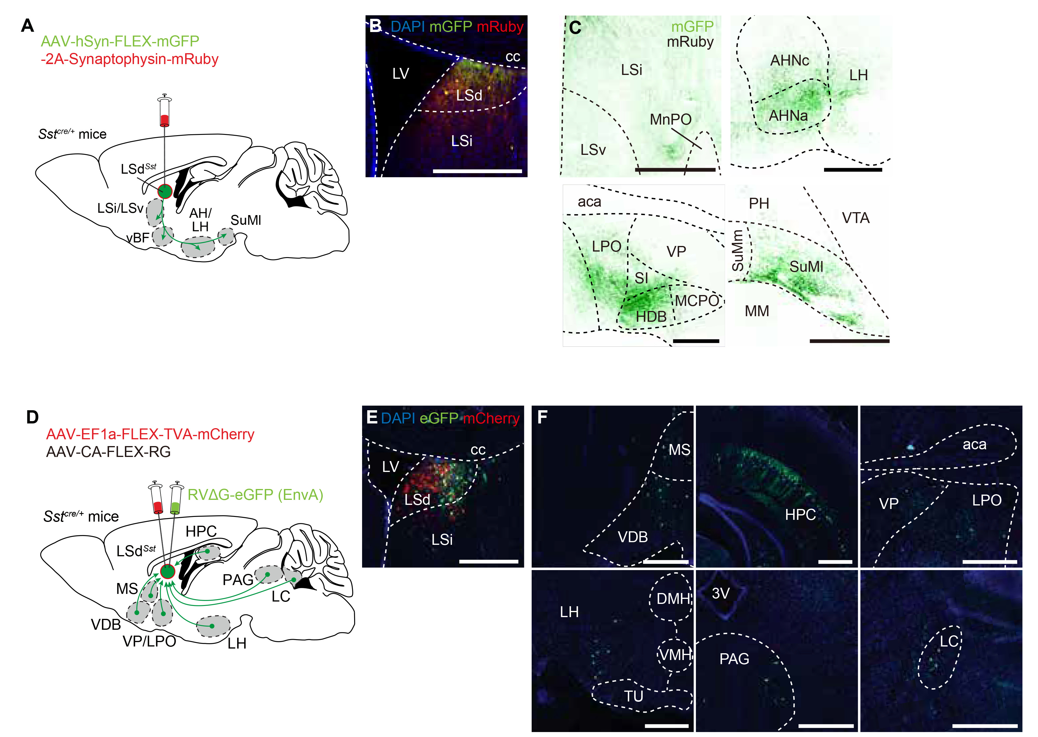
Fig. 2. LSdSst neurons are connected with regions implicated in stress response. (A) AAV vector expressing cytosol-filling mGFP and terminal bouton-labeling mRuby-fused synaptophysin was injected into the LSd of the Sstcre /+ mice for anterograde tracing (n=3). Summary of the tracing experiment is shown. (B) Example image of the injection site, showing the expression of both mRuby and mGFP. (C) Representative images of the identified projection targets. Green, mGFP; black, mRuby. MnPO, median preoptic area; AH, anterior hypothalamus; SI, substantia innominata; HDB, horizontal limb of the diagonal band of Broca; MCPO, magnocellular preoptic nucleus; PH, posterior hypothalamus; VTA, ventral tegmental area; SuMm, medial supramammillary nucleus; SuMl, lateral supramammillary nucleus; MM, medial mammillary nucleus. Scale bars, 500 μm (C), 50 μm (B, insets). (D) AAV vectors Cre-dependently expressing rabies G protein and TVA receptor, and G-deficient EnvA-pseudotyped RV carrying eGFP were injected into the LSd of the Sstcre /+ mice for tracing monosynaptic inputs of LSdSst neurons (n=3). Summary of the retrograde tracing is shown. (E) Representative confocal image of the injection site, showing the expression of mCherry fused to TVA (red) and eGFP (green). (F) Representative images of monosynaptically connected upstream neurons of LSdSst neurons. MS, medial septum; VDB, nucleus of the vertical limb of the diagonal band; HPC, hippocampus; aca, anterior commissure; VP, ventral pallidum; LPO, lateral preoptic area; LH, lateral hypothalamus; DMH, dorsomedial hypothalamic nucleus; VMH, ventromedial hypothalamic nucleus; TU, tuberal nucleus; 3V, third ventricle; PAG, periaqueductal gray; LC, locus coeruleus.
© Exp Neurobiol