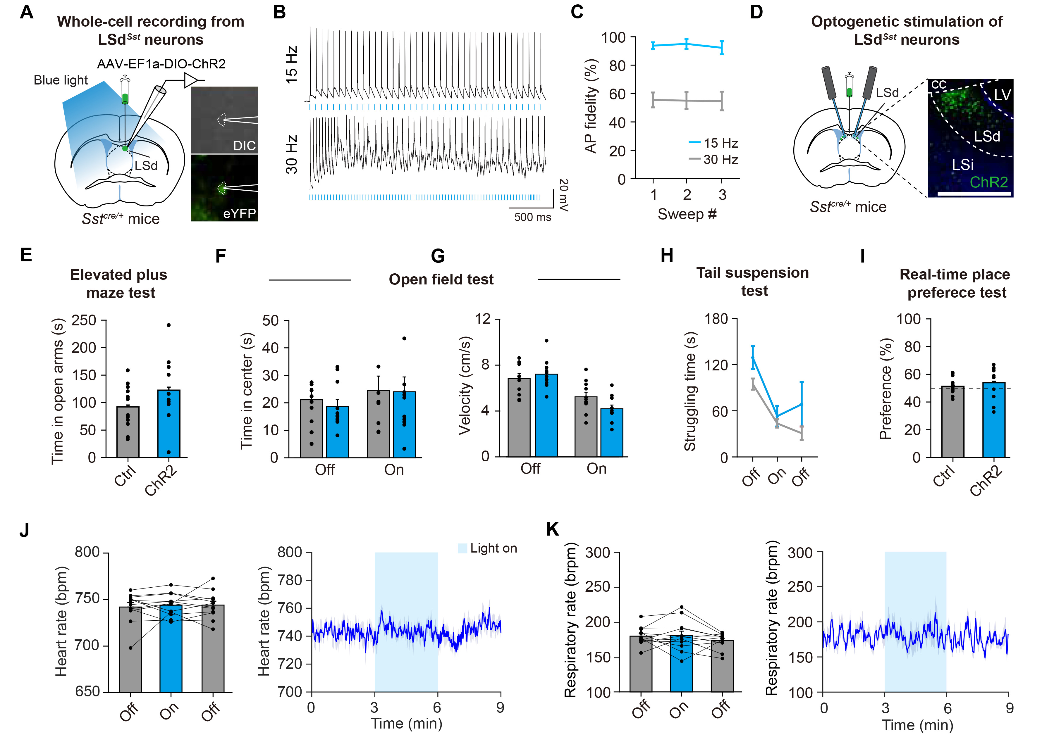
Fig. 4. Optogenetic stimulation of LSdSst neurons does not affect stress-related behaviors or autonomic functions. (A) Schematic of patch-clamp recording from ChR2-expressing LSdSst neurons. The LS area was illuminated with blue light for optogenetic stimulations. Right, example images of a LSdSst neuron expressing ChR2-eYFP during recording. DIC, differential interference contrast. (B) Representative traces showing the responses of LSdSst neurons upon the delivery of light pulse trains in 15 Hz or 30 Hz. (C) 15 Hz blue laser-stimulation evokes action potentials (AP) with high fidelity, whereas 30 Hz stimulation shows 50~60% of success rate in inducing action potentials (n=4 cells from 2 animals). (D) Schematics showing Cre-dependent expression of ChR2 and fiberoptic cannula implantation for bilateral optogenetic stimulations of LSdSst. Representative confocal image shows LSd-restricted expression of ChR2. Scale bar, 200 µm. (E~K) Optogenetic stimulation of LSdSst neurons did not affect time spent in open arms of the elevated plus maze (n=11 ChR2, n=12 Ctrl, p=0.339) (E), time spent in the center (n=11 ChR2, n=11 Ctrl, two-way repeated measures ANOVA interaction, F(1,20)=0.097, p=0.759) (F) or velocity (n=11 ChR2, n=11 Ctrl, two-way repeated measures ANOVA interaction, F(1,20)=7.787, p=0.011) (G) in an open field arena, struggling time in the tail suspension test (n=4 ChR2, n=7 Ctrl, two-way repeated measures ANOVA interaction, F(2,18)=0.778, p=0.474) (H), nor real-time place preference (n=11 ChR2, n=12 Ctrl, p=0.347) (I). Dashed line indicates 50% preference. This manipulation also did not affect heart rate (n=12, one-way repeated measures ANOVA interaction, F(2,22)=0.199, p=0.821) (J) and respiratory rate (n=12, one-way repeated measures ANOVA interaction, F(2,22)=1.114, p=0.339) (K), Data are represented as mean±s.e.m. Asterisks indicate significance levels for comparisons in each panel using Bonferroni post-tests following two-way repeated measures ANOVA or Wilcoxon rank-sum test.
© Exp Neurobiol