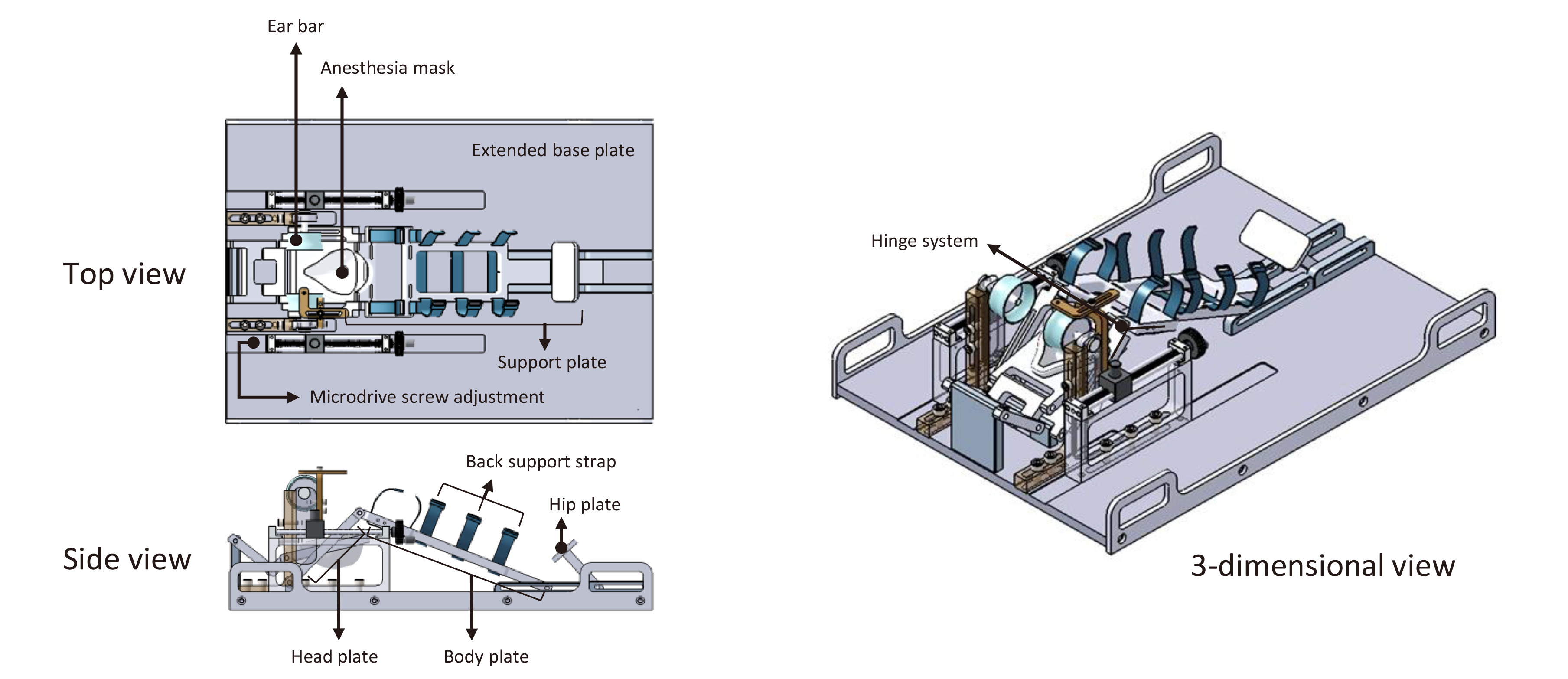
Fig. 2. Appearance of the custom-built CT-MRI compatible stereotaxic frame. The customized stereotaxic frame showing the top, side, and three-dimensional view. Top view shows the position of the extended base plate, the microdrive screw adjustment for the anteroposterior axis, anesthesia mask, ear bar, and support plate. Anteroposterior adjustment for adaptor is 150 mm. Side view shows the back strap and the support plates including head, body and hip for prone position. Three-dimensional view shows the hinge system, which are angle adjustable to stabilize and maintain the flexion of upper cervical spine.
© Exp Neurobiol