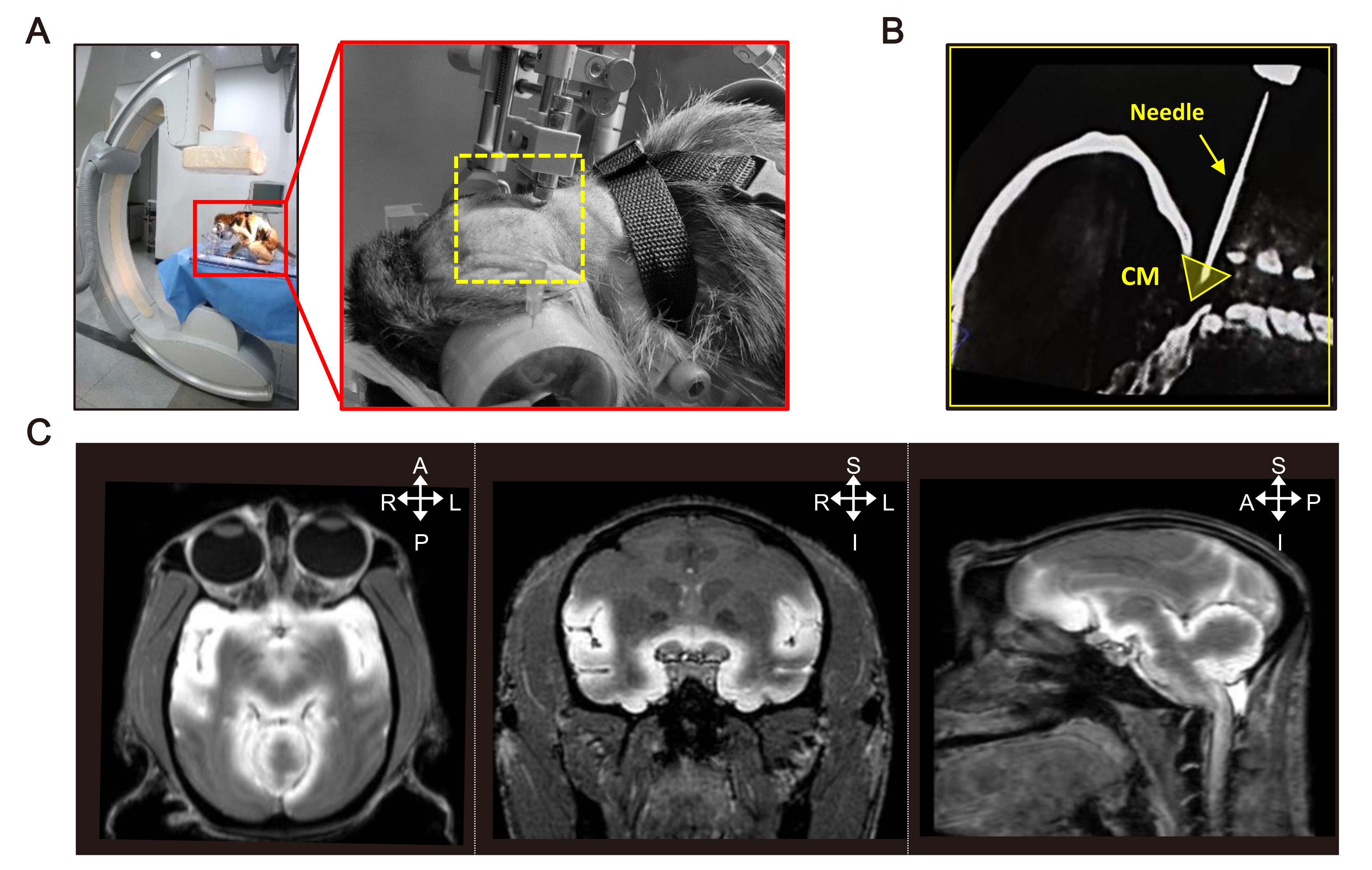
Fig. 3. Application of the custom-built CT-MRI compatible stereotaxic frame. (A) Picture of a monkey anesthetized and immobilized within a stereotaxic frame for XperCT scanning using the flat detector C-arm. (B) Representative XperCT image showing the accuracy and precision of the injection into the cisterna magna target region. (C) Representative MRI images obtained after the CSF tracer injection into the cisterna magna. The CSF tracer used is a gadolinium-based MR contrast agent.
© Exp Neurobiol