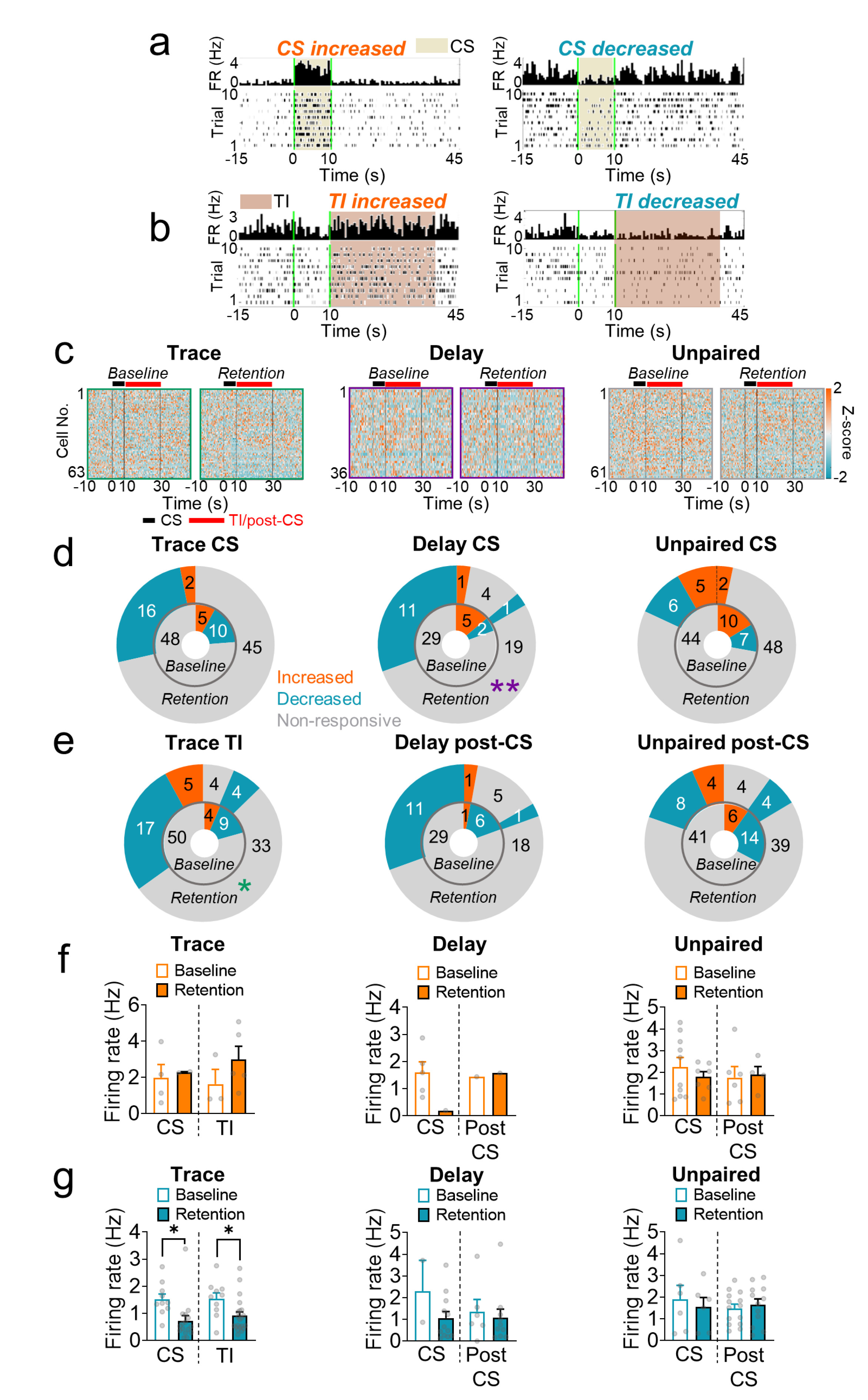
Fig. 3. Activity of EC units during the CS and TI (post-CS) from Trace, Delay, and Unpaired. (a) The peri-event histograms (PETHs) and raster plots of representative EC cells that showed increased firing rates during the CS (left) and decreased firing rates during the CS (right). (b) The PETHs and raster plots of representative EC cells that showed increased firing rates during the TI (left) and decreased firing rates during the TI (right). (c) Z-scored heatmaps of cells during baseline and retention from Trace (left), Delay (middle), and Unpaired (right). Each row indicates firing rates of individual cells throughout the baseline and retention periods. (d) The proportion of CS-responsive cells during the baseline (inner donut chart) and the retention (outer donut chart) sessions from Trace (left), Delay (middle), and Unpaired (right). (e) The proportion of TI (post-CS)-responsive cells during the baseline (inner donut chart) and the retention (outer donut chart) sessions from Trace (left), Delay (middle), and Unpaired (right). (f) The mean firing rates of the CS-increased cells from the baseline vs. retention sessions and the mean firing rates of the TI (post-CS)-increased cells from the baseline vs. retention sessions (left, Trace; middle, Delay; right, Unpaired). (g) The mean firing rates of the CS-decreased cells from the baseline vs. retention sessions and the mean firing rates of the TI (post-CS)-decreased cells from the baseline vs. retention sessions (left, Trace; middle, Delay; right, Unpaired). Data for e and f were assessed by paired t-test (*p<0.05), and each circle represents individual cell data. All data are represented as mean±SEM.
© Exp Neurobiol