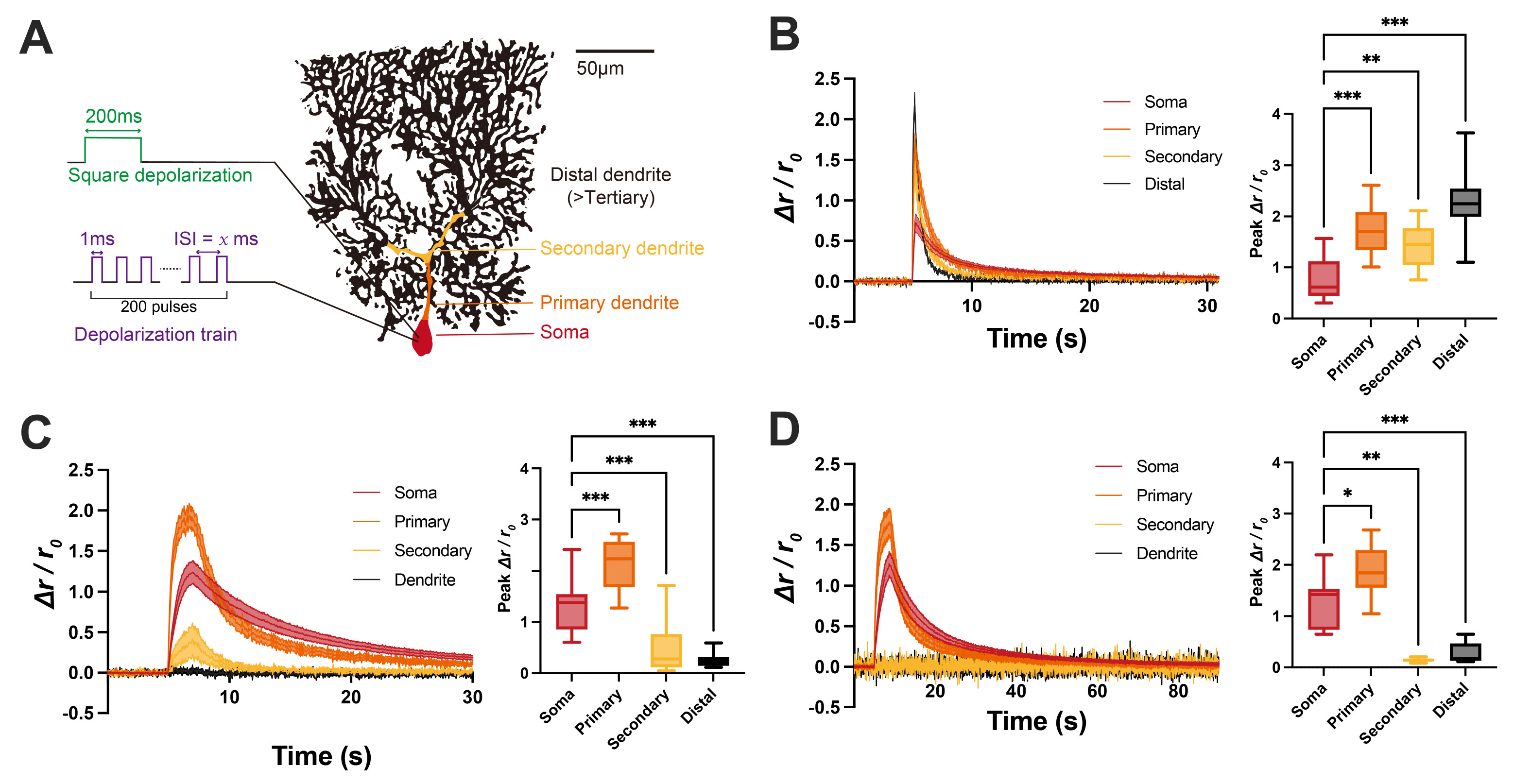
Fig. 1. Ca2+ propagation in Purkinje cells under natural simple spike firing. (A) Schematic figure for experiment and regional separation of Purkinje cells. (B) Ca2+ transients triggered by 200 ms square depolarization. The peak Δr/r0 of soma (n=13) was significantly lower than all dendritic regions (Primary, n=12, p<0.001; Secondary, n=9, p=0.007; Distal, n=30, p<0.001). (C) Ca2+ transients induced by 1ms depolarization train with 9 ms interspike interval (ISI). The peak Δr/r0 of soma (n=13) was significantly lower than all dendritic regions primary dendrite (n=12, p<0.001). However, the secondary and distal dendrites showed considerably lower transients than soma (Secondary, n=9, p<0.001; Distal, n=28, p<0.001). (D) Ca2+ transients induced by 1 ms depolarization train with 19 ms interval. Similar to the 9 ms interval results, the peak Δr/r0 of soma (n=11) was significantly lower than Primary dendrite (n=9, p=0.013), but there were no Ca2+ transients in the secondary (n=2, p=0.006) and distal dendrites (n=7, p<0.001). One-way ANOVA with post-hoc Dunnett test was used for multiple comparisons. Error bar indicates SEM. *p<0.05, **p<0.01, ***p<0.001.
© Exp Neurobiol