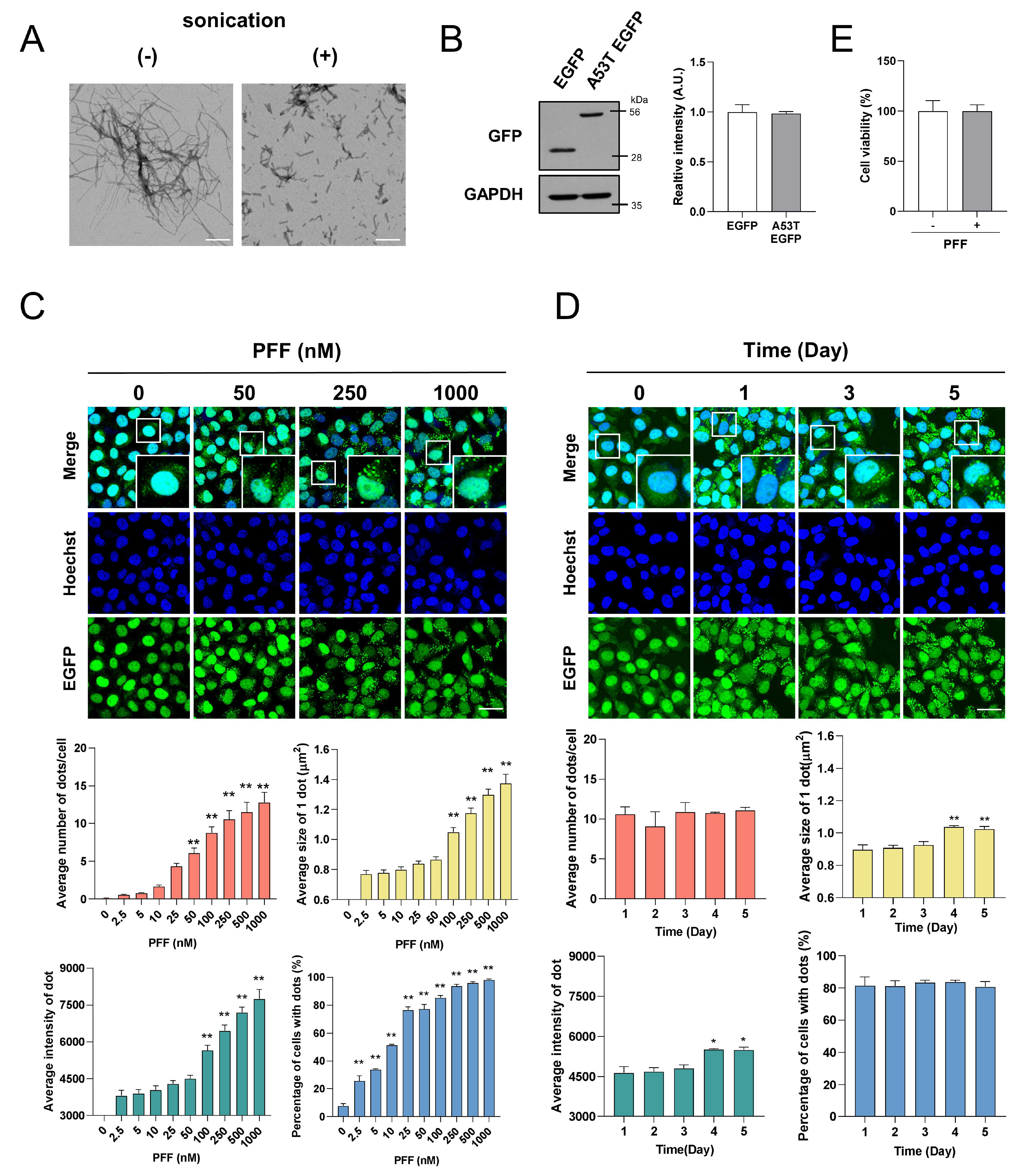
Fig. 1. Exogenously added PFF induces aggregation puncta consisting of A53T-α-syn-EGFP in a time- and dose-dependent manner. (A) After sonication, PFF were visualized with transmission electron microscopy. Scale bars indicate 200 nm. (B) EGFP and A53T α-syn-EGFP SH-SY5Y cells were lyzed with lysis buffer, then western blot for EGFP was performed. A53T α-syn-EGFP SH-SY5Y cells were incubated with indicated doses of PFF for 3 days (C) or with 250 nM PFF for indicated times (D). Then, the samples were observed under a confocal microscopy. (E) Cytotoxicity assay was performed after 5 days of treatment with 1 μM PFF. The values were derived from more than 3 independent experiments. Blue indicates Hoechst staining. Scale bars indicate 50 μm. *p<0.05, **p<0.01 against control, one-way ANOVA.
© Exp Neurobiol