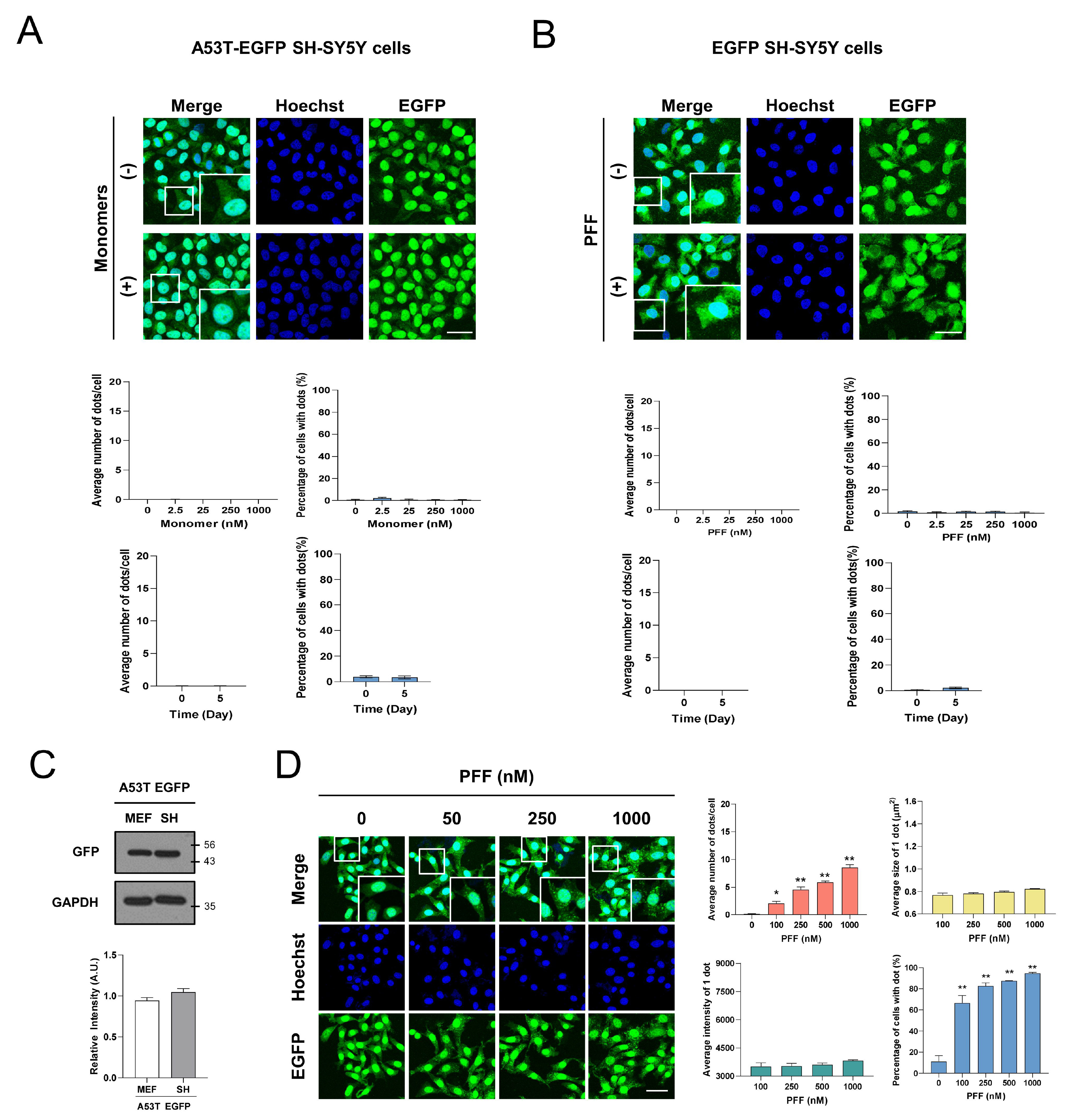
Fig. 2. Aggregation puncta of A53T α-syn-EGFP induced by PFF were formed in α-syn species- and A53T α-syn-EGFP-specific manners. (A) A53T α-syn-EGFP SH-SY5Y cells were incubated with indicated doses of α-syn monomers for 1 day or with 250 nM α-syn monomers for 5 days. (B) EGFP SH-SY5Y cells were incubated with indicated doses of PFF for 1 day or with 250 nM PFF for 5 days. (C) A53T α-syn-EGFP MEF cells and A53T α-syn-EGFP SH-SY5Y cells were lyzed with lysis buffer, then western blot for EGFP was performed. (D) A53T α-syn-EGFP MEF cells were incubated with indicated doses of PFF for 1 day. The values were derived from 3 independent experiments. Blue indicates Hoechst staining. Scale bars indicate 50 μm. *p<0.05, **p<0.01 against control, unpaired t-test or one-way ANOVA.
© Exp Neurobiol