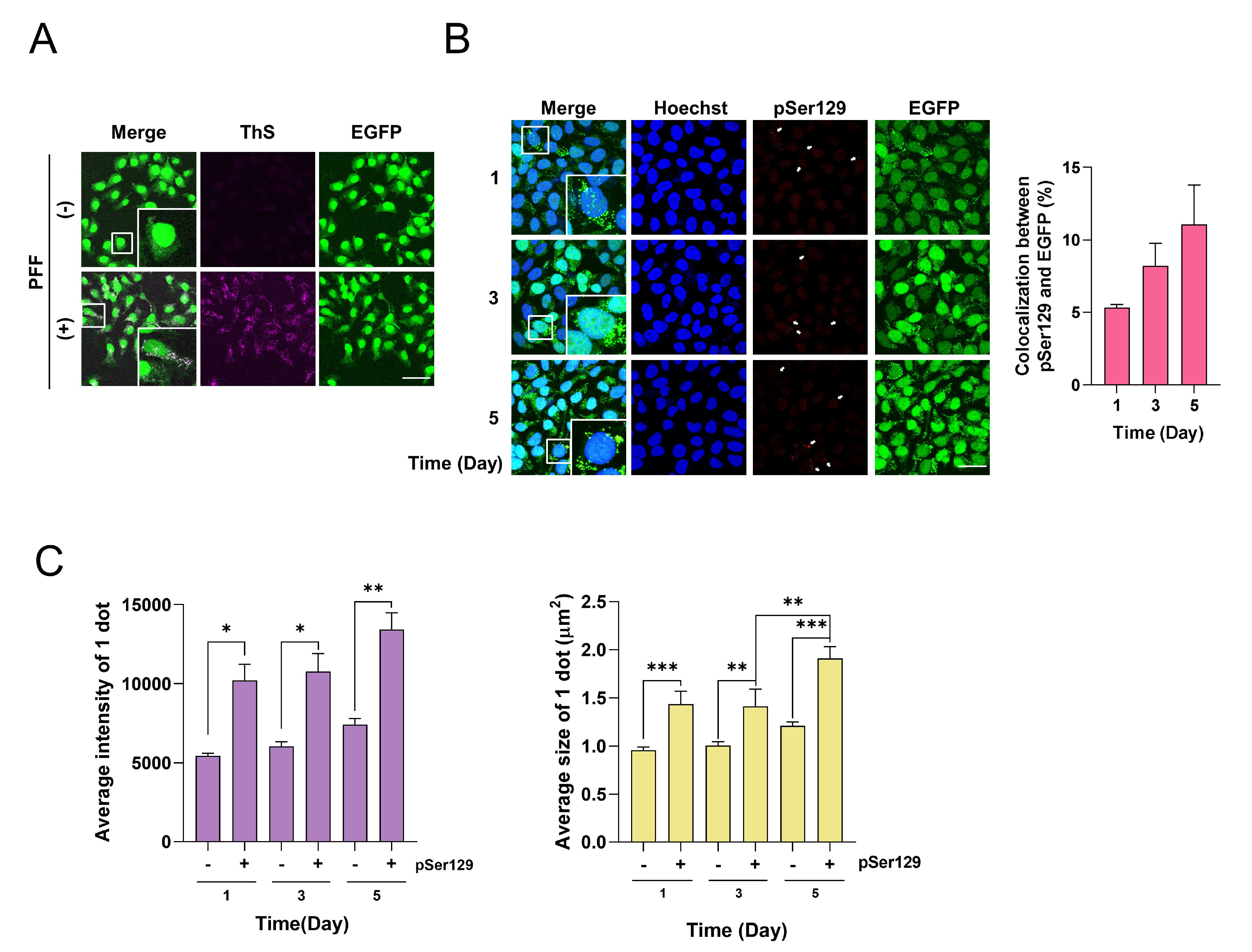
Fig. 3. Inclusions of A53T α-syn-EGFP contain pSer129 α-syn in a time-dependent manner. (A) A53T α-syn-EGFP SH-SY5Y cells were incubated with 250 nM PFF for 1 day. Then, the samples were stained with 0.025% thioflavin S (ThS, Violet). (B) A53T α-syn-EGFP SH-SY5Y cells were incubated with 250 nM PFF for 1 day. Then, the samples were stained with pSer129 α-syn antibody (Red). The values were derived from 3 independent experiments. The samples were observed under a confocal microscopy. Blue indicates Hoechst staining. Scale bars indicate 50 μm. (C) The intensity of the dots and their size were analyzed after dividing them into those that were colocalized with pSer129 α-syn or not. *p<0.05, **p<0.01, ***p<0.001 against control, one-way ANOVA.
© Exp Neurobiol