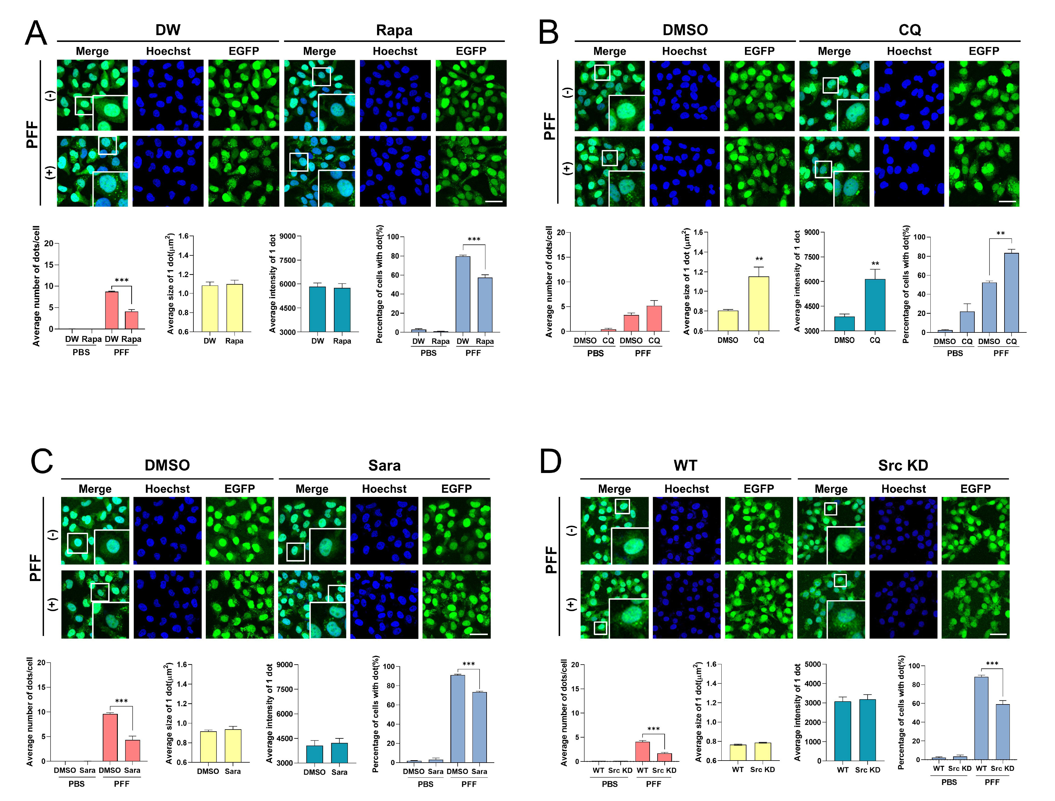
Fig. 4. Pharmacologically and genetically modifications altered the inclusion body formation of A53T α-syn-EGFP induced by PFF. A53T α-syn-EGFP SH-SY5Y cells were incubated with 250 nM PFF in the presence or absence of 10 μM rapamycin (Rapa) (A) or 40 μM chloroquine (CQ) (B), for 1 day. (C) A53T α-syn-EGFP SH-SY5Y cells were incubated with 250 nM PFF in the presence or absence of 1 μM saracatinib (Sara) for 1 day. (D) A53T α-syn-EGFP/c-src KD SH-SY5Y cells were incubated with 250 nM PFF for 1 day. The samples were observed under a confocal microscopy. The values were derived from 3 independent experiments. Blue indicates Hoechst staining. Scale bars indicate 50 μm. **p<0.01, ***p<0.001 against control, unpaired t-test or one-way ANOVA.
© Exp Neurobiol