Articles
Article Tools
Stats or Metrics
Article
Original Article
Exp Neurobiol 2019; 28(6): 670-678
Published online December 31, 2019
https://doi.org/10.5607/en.2019.28.6.670
© The Korean Society for Brain and Neural Sciences
The Molecular Signatures of Acute-immobilization-induced Antinociception and Chronic-immobilization-induced Antinociceptive Tolerance
Jing-Hui Feng, Hee-Jung Lee and Hong-Won Suh*
Department of Pharmacology and Institute of Natural Medicine, College of Medicine, Hallym University, Chuncheon 24252, Korea
Correspondence to: *To whom correspondence should be addressed.
TEL: 82-33-248-2614, FAX: 82-33-248-2612
e-mail: hwsuh@hallym.ac.kr.
This is an Open Access article distributed under the terms of the Creative Commons Attribution Non-Commercial License(http://creativecommons.org/licenses/by-nc/4.0) which permits unrestricted non-commercial use, distribution, andreproduction in any medium, provided the original work is properly cited.
Abstract
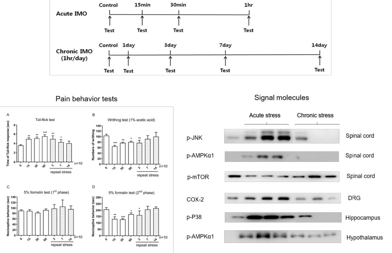
In the present study, the productions of antinociception induced by acute and chronic immobilization stress were compared in several animal pain models. In the acute immobilization stress model (up to 1 hr immobilization), the antinociception was produced in writhing, tail-flick, and formalin-induced pain models. In chronic immobilization stress experiment, the mouse was enforced into immobilization for 1 hr/day for 3, 7, or 14 days, then analgesic tests were performed. The antinociceptive effect was gradually reduced after 3, 7 and 14 days of immobilization stress. To delineate the molecular mechanism involved in the antinociceptive tolerance development in the chronic stress model, the expressions of some signal molecules in dorsal root ganglia (DRG), spinal cord, hippocampus, and the hypothalamus were observed in acute and chronic immobilization models. The COX-2 in DRG, p-JNK, p-AMPKα1, and p-mTOR in the spinal cord, p-P38 in the hippocampus, and p-AMPKα1 in the hypothalamus were elevated in acute immobilization stress, but were reduced gradually after 3, 7 and 14 days of immobilization stress. Our results suggest that the chronic immobilization stress causes development of tolerance to the antinociception induced by acute immobilization stress. In addition, the COX-2 in DRG, p-JNK, p-AMPKα1, and p-mTOR in the spinal cord, p-P38 in the hippocampus, and p-AMPKα1 in the hypothalamus may play important roles in the regulation of antinociception induced by acute immobilization stress and the tolerance development induced by chronic immobilization stress.
Graphical Abstract

Keywords: Acute stress, Chronic stress, Antinociception, Tolerance, Signal molecule


