Articles
Article Tools
Supplementary
Stats or Metrics
Article
Original Article
Exp Neurobiol 2020; 29(1): 80-92
Published online February 29, 2020
https://doi.org/10.5607/en.2020.29.1.80
© The Korean Society for Brain and Neural Sciences
Enhanced Bidirectional Connectivity of the Subthalamo-pallidal Pathway in 6-OHDA-mouse Model of Parkinson's Disease Revealed by Probabilistic Tractography of Diffusion-weighted MRI at 9.4T
A-Yoon Kim1, Chiwoo Oh2, Hyung-Jun Im2 and Hyeon-Man Baek1,3*
1Department of Health Science and Technology, GAIHST, Gachon University, Incheon 21936, 2Department of Transdisciplinary Studies, Graduate School of Convergence Science and Technology, Seoul National University, Seoul 16229, 3Lee Gil Ya Cancer & Diabetes Institute, Gachon University, Incheon 21999, Korea
Correspondence to: *To whom correspondence should be addressed.
TEL: 82-32-899-6678, FAX: 82-32-899-6677
e-mail: hmbaek98@gachon.ac.kr
This is an Open Access article distributed under the terms of the Creative Commons Attribution Non-Commercial License(http://creativecommons.org/licenses/by-nc/4.0) which permits unrestricted non-commercial use, distribution, andreproduction in any medium, provided the original work is properly cited.
Abstract
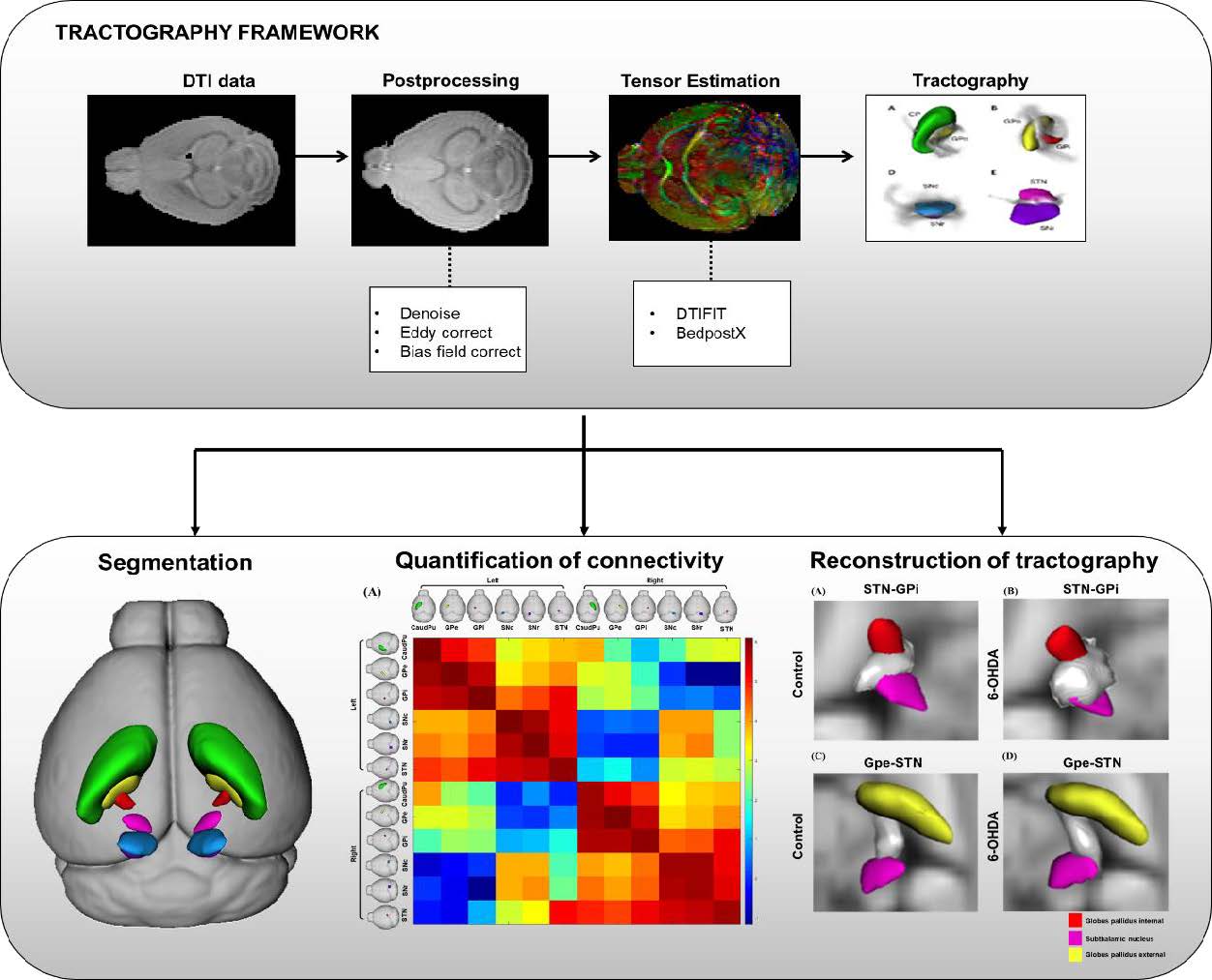
An important challenge in Parkinson’s disease (PD) based neuroscience and neuroimaging is mapping the neuronal connectivity of the basal ganglia to understand how the disease affects brain circuitry. However, a majority of diffusion tractography studies have shown difficulties in revealing connections between distant anatomic brain regions and visualizing basal ganglia connectome. In this current study, we investigated the differences in basal ganglia connectivity between 6-OHDA induced
Graphical Abstract

Keywords: Basal ganglia, Parkinson’s disease, Diffusion MRI, Probabilistic tractography, Globus pallidus, Subthalamic nucleus


