Articles
Article Tools
Stats or Metrics
Article
Original Article
Exp Neurobiol 2020; 29(3): 249-272
Published online June 30, 2020
https://doi.org/10.5607/en20019
© The Korean Society for Brain and Neural Sciences
Pim-1 Protects Retinal Ganglion Cells by Enhancing Their Regenerative Ability Following Optic Nerve Crush
Shoumei Zhang1,4, Li Shuai2, Dong Wang1, Tingting Huang1, Shengsheng Yang3, Mingyong Miao3, Fang Liu1 and Jiajun Xu1*
1Department of Anatomy, Second Military Medical University, Shanghai 200433, 2Department of Health Administration, Second Military Medical University, Shanghai 200433, 3Department of Biochemistry and Molecular Biology, Second Military Medical University, Shanghai 200433, 4Translational Medical Center for Stem Cell Therapy, Shanghai East Hospital, Tongji University School of Medicine, Shanghai 200120, China
Correspondence to: *To whom correspondence should be addressed.
TEL: 86-21-81870950, FAX: 86-21-81870955
e-mail: xujiajunsmmu@163.com
This is an Open Access article distributed under the terms of the Creative Commons Attribution Non-Commercial License
(http://creativecommons.org/licenses/by-nc/4.0) which permits unrestricted non-commercial use, distribution, and
reproduction in any medium, provided the original work is properly cited.
Abstract
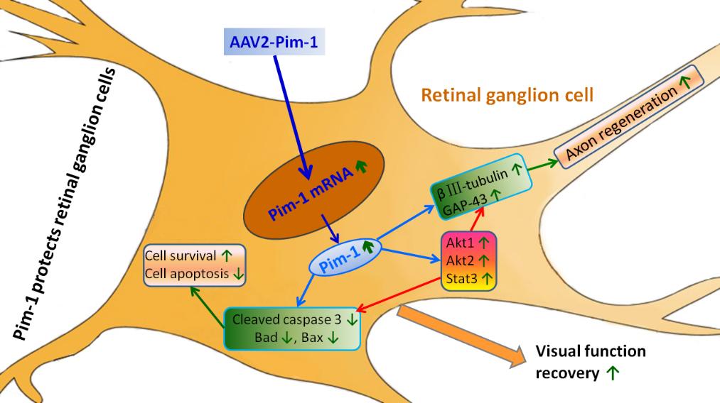
Provirus integration site Moloney murine leukemia virus (Pim-1) is a proto-oncogene reported to be associated with cell proliferation, differentiation and survival. This study was to explore the neuroprotective role of Pim-1 in a rat model subjected to optic nerve crush (ONC), and discuss its related molecules in improving the intrinsic regeneration ability of retinal ganglion cells (RGCs). Immunofluorescence staining showed that AAV2- Pim-1 infected 71% RGCs and some amacrine cells in the retina. Real-time PCR and Western blotting showed that retina infection with AAV2- Pim-1 up-regulated the Pim-1 mRNA and protein expressions compared with AAV2-GFP group. Hematoxylin-Eosin (HE) staining, γ-synuclein immunohistochemistry, Cholera toxin B (CTB) tracing and TUNEL showed that RGCs transduction with AAV2-Pim-1 prior to ONC promoted the survival of damaged RGCs and decreased cell apoptosis. RITC anterograde labeling showed that Pim-1 overexpression increased axon regeneration and promoted the recovery of visual function by pupillary light reflex and flash visual evoked potential. Western blotting showed that Pim- 1 overexpression up-regulated the expression of Stat3, p-Stat3, Akt1, p-Akt1, Akt2 and p-Akt2, as well as βIII-tubulin, GAP-43 and 4E-BP1, and downregulated the expression of SOCS1 and SOCS3, Cleaved caspase 3, Bad and Bax. These results demonstrate that Pim-1 exerted a neuroprotective effect by promoting nerve regeneration and functional recovery of RGCs. In addition, it enhanced the intrinsic regeneration capacity of RGCs after ONC by activating Stat3, Akt1 and Akt2 pathways, and inhibiting the mitochondrial apoptosis pathways. These findings suggest that Pim-1 may prove to be a potential therapeutic target for the clinical treatment of optic nerve injury.
Graphical Abstract

Keywords: Pim-1, Nerve regeneration, Retinal ganglion cell, Optic nerve injury


