Articles
Article Tools
Stats or Metrics
Article
Original Article
Exp Neurobiol 2021; 30(4): 294-307
Published online August 31, 2021
https://doi.org/10.5607/en21014
© The Korean Society for Brain and Neural Sciences
Hyperoxygenation Treatment Reduces Beta-amyloid Deposition via MeCP2-dependent Upregulation of MMP-2 and MMP-9 in the Hippocampus of Tg-APP/PS1 Mice
Juli Choi1, Hyejin Kwon1 and Pyung-Lim Han1,2*
1Departments of Brain and Cognitive Sciences and 2Chemistry and Nano Science, Ewha Womans University, Seoul 03760, Korea
Correspondence to: *To whom correspondence should be addressed.
TEL: 82-2-3277-4130, FAX: 82-2-3277-3419
e-mail: plhan@ewha.ac.kr
This is an Open Access article distributed under the terms of the Creative Commons Attribution Non-Commercial License (http://creativecommons.org/licenses/by-nc/4.0) which permits unrestricted non-commercial use, distribution, and reproduction in any medium, provided the original work is properly cited.
Abstract
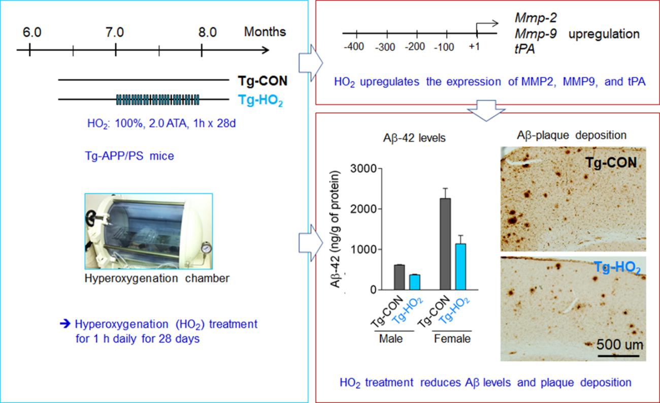
Recently we reported that hyperoxygenation treatment reduces amyloid-beta accumulation and rescues cognitive impairment in the Tg-APP/PS1 mouse model of Alzheimer’s disease. In the present study, we continue to investigate the mechanism by which hyperoxygenation reduces amyloid-beta deposition in the brain. Hyperoxygenation treatment induces upregulation of matrix metalloproteinase-2 (MMP-2), MMP-9, and tissue plasminogen activator (tPA), the endopeptidases that can degrade amyloid-beta, in the hippocampus of Tg-APP/PS1 mice. The promoter regions of the three proteinase genes all contain potential binding sites for MeCP2 and Pea3, which are upregulated in the hippocampus after hyperoxygenation. Hyperoxygenation treatment in HT22 neuronal cells increases MeCP2 but not Pea3 expression. In HT22 cells, siRNA-mediated knockdown of
Graphical Abstract

Keywords: Hyperoxygenation, Beta-amyloid, MeCP2, MMP-2, MMP-9


