Articles

  • KSBNS 2024

Article

Original Article

Exp Neurobiol 2018; 27(6): 508-525

Published online December 28, 2018

https://doi.org/10.5607/en.2018.27.6.508

© The Korean Society for Brain and Neural Sciences

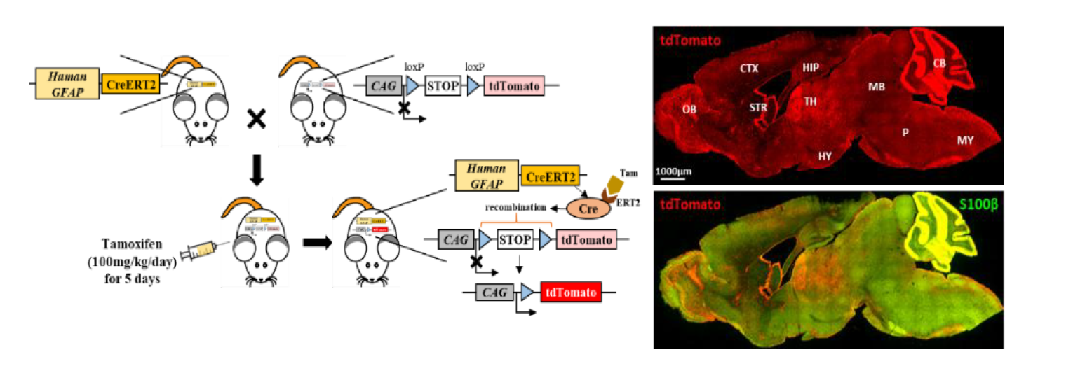
Astrocyte Specificity and Coverage of hGFAP-CreERT2 [Tg(GFAP-Cre/ERT2)13Kdmc] Mouse Line in Various Brain Regions

Yongmin Mason Park1,2,3,†, Heejung Chun2,3,†, Jeong-Im Shin1,2,3, and C. Justin Lee1,2,3*

1Division of Bio-Medical Science & Technology, Department of Neuroscience, KIST School, Korea University of Science and Technology, Seoul 02792, Korea.

2Center for Glia-Neuron Interaction, Korea Institute of Science and Technology (KIST), Seoul 02792, Korea.

3Center for Cognition and Sociality, Institute for Basic Science, Daejeon 34126, Korea.

Correspondence to: *To whom correspondence should be addressed.
TEL: 82-42-878-9150, FAX: 82-42-878-9151
e-mail: cjl@ibs.re.kr
These authors contributed equally to this work.

Received: November 30, 2018; Revised: December 12, 2018; Accepted: December 12, 2018

Astrocyte is the most abundant cell type in the central nervous system and its importance has been increasingly recognized in the brain pathophysiology. To study in vivo function of astrocyte, astrocyte-specific gene-targeting is regarded as a powerful approach. Especially, hGFAP-CreERT2, which expresses tamoxifen-inducible Cre recombinase under the human GFAP promoter, has been developed and characterized from several research groups. However, one of these mouse lines, [Tg(GFAP-Cre/ERT2)13Kdmc] from Ken McCarthy group has not been quantitatively analyzed, despite its frequent use. Here, we performed comprehensive characterization of this mouse line with quantitative analysis. By crossing this mouse line with Ai14 (RCL-tdTomato), a very sensitive Cre reporter mouse line, we visualized the Cre-expressing cells in various brain regions. For quantitative analysis, we immunostained S100β as an astrocytic marker and NeuN, tyrosine hydroxylase or calbindin as a neuronal marker in different brain regions. We calculated ‘astrocyte specificity’ as the proportion of co-labelled S100β and tdTomato positive cells in the total number of tdTomato positive cells and the ‘astrocyte coverage’ as the proportion of co-labelled S100β and tdTomato positive cells in the total number of S100β positive cells. Interestingly, we found varying degree of astrocyte specificity and coverage in each brain region. In cortex, hypothalamus, substantia nigra pars compacta and cerebellar Purkinje layer, we observed high astrocyte specificity (over 89%) and relatively high astrocyte coverage (over 70%). In striatum, hippocampal CA1 layer, dentate gyrus and cerebellar granule layer, we observed high astrocyte specificity (over 80%), but relative low astrocyte coverage (50–60%). However, thalamus and amygdala showed low astrocyte specificity (about 65%) and significant neuron specificity (over 30%). This hGFAP-CreERT2 mouse line can be useful for genetic modulations of target gene either in gain-of-function or loss-of-function studies in the brain regions with high astrocyte specificity and coverage. However, the use of this mouse line should be restricted to gain-of-function studies in the brain regions with high astrocyte specificity but low coverage. In conclusion, hGFAP-CreERT2 mouse line could be a powerful tool for gene-targeting of astrocytes in cortex, striatum, hippocampus, hypothalamus, substantia nigra pars compacta and cerebellum, but not in thalamus and amygdala.

Graphical Abstract


Keywords: Astrocytes, Glial fibrillary astrocytic protein, Cre recombinase, Tamoxfien