Articles

Article Tools
 View Full Text View Full Text |
 Abstract Abstract |
 Article as PDF Article as PDF |
 Print this Article Print this Article |
 Pubmed Pubmed |
 PMC PMC |
 PubReader PubReader |
 Export to Citation Export to Citation |
 Email Alerts Email Alerts |
 Open Access Open Access |
Share this article on :


Stats or Metrics
Article
Original Article
Exp Neurobiol 2019; 28(3): 362-375
Published online June 26, 2019
https://doi.org/10.5607/en.2019.28.3.362
© The Korean Society for Brain and Neural Sciences
Quantitative Proteomic Analysis Reveals Impaired Axonal Guidance Signaling in Human Postmortem Brain Tissues of Chronic Traumatic Encephalopathy
Baibin Bi1,2,3,†, Han-Pil Choi4,†, Seung Jae Hyeon5,†, Shengnan Sun2, Ning Su1, Yuguang Liu3, Junghee Lee1,4,6, Neil W. Kowall1,4,6, Ann C. McKee1,4,6*, Jing-Hua Yang1,2,4*, and Hoon Ryu1,4,5,6*
1Departments of Neurology, Pathology, and Surgery, Boston University School of Medicine, Boston, MA 02118, USA.
2Cancer Research Center, Shandong University School of Medicine, Jinan 250012, China.
3Department of Neurosurgery, Qilu Hospital of Shandong University, Jinan 250012, China.
4Proteomics Laboratory, VA Boston Healthcare System, Boston, MA 02130, USA.
5Center for Neuromedicine, Brain Science Institute, Korea Institute of Science and Technology, Seoul 04535, Korea.
6Boston University Alzheimer's Disease Center (BU ADC) and Chronic Traumatic Encephalopathy (CTE) Center, Boston University School of Medicine, Boston, MA 02118, USA.
Correspondence to: *To whom correspondence should be addressed.
Ann C. McKee, TEL: 1-857-364-5707, FAX: 1-857-364-4540, e-mail: amckee@bu.edu
Jing-Hua Yang, TEL: 1-857-364-5611, FAX: 1-857-364-5627, e-mail: jyang@bu.edu
Hoon Ryu, TEL: 1-857-364-5910, FAX: 1-857-364-4540, e-mail: hoonryu@bu.edu
†These authors are equally contributed.
Abstract
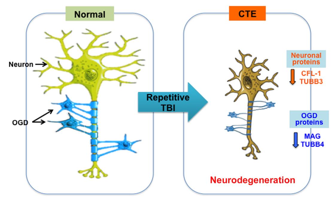
Chronic traumatic encephalopathy (CTE) is a distinct neurodegenerative disease that associated with repetitive head trauma. CTE is neuropathologically defined by the perivascular accumulation of abnormally phosphorylated tau protein in the depths of the sulci in the cerebral cortices. In advanced CTE, hyperphosphorylated tau protein deposits are found in widespread regions of brain, however the mechanisms of the progressive neurodegeneration in CTE are not fully understood. In order to identify which proteomic signatures are associated with CTE, we prepared RIPA-soluble fractions and performed quantitative proteomic analysis of postmortem brain tissue from individuals neuropathologically diagnosed with CTE. We found that axonal guidance signaling pathwayrelated proteins were most significantly decreased in CTE. Immunohistochemistry and Western blot analysis showed that axonal signaling pathway-related proteins were down regulated in neurons and oligodendrocytes and neuron-specific cytoskeletal proteins such as TUBB3 and CFL1 were reduced in the neuropils and cell body in CTE. Moreover, oligodendrocyte-specific proteins such as MAG and TUBB4 were decreased in the neuropils in both gray matter and white matter in CTE, which correlated with the degree of axonal injury and degeneration. Our findings indicate that deregulation of axonal guidance proteins in neurons and oligodendrocytes is associated with the neuropathology in CTE. Together, altered axonal guidance proteins may be potential pathological markers for CTE.
Graphical Abstract
Keywords: Chronic traumatic encephalopathy (CTE), Quantitative proteomics, Axonal guidance, Oligodendrocyte, Neuron, Neurodegeneration