Articles

Article Tools
 View Full Text View Full Text |
 Abstract Abstract |
 Article as PDF Article as PDF |
 Print this Article Print this Article |
 Pubmed Pubmed |
 PMC PMC |
 PubReader PubReader |
 Export to Citation Export to Citation |
 Email Alerts Email Alerts |
 Open Access Open Access |
Supplementary
Share this article on :


Stats or Metrics
Article
Original Article
Exp Neurobiol 2021; 30(2): 183-202
Published online April 30, 2021
https://doi.org/10.5607/en20056
© The Korean Society for Brain and Neural Sciences
Preliminary Comparison of Subcortical Structures in Elderly Subclinical Depression: Structural Analysis with 3T MRI
SangJin Im1, Jeonghwan Lee2 and Siekyeong Kim2*
1Lee Gil Ya Cancer & Diabetes Institute, Gachon University, Incheon 21999,
2Department of Psychiatry, Chungbuk National University College of Medicine, Cheongju 28644, Korea
Correspondence to: *To whom correspondence should be addressed.
TEL: 82-43-269-6364, FAX: 82-43-267-7951
e-mail: poshong@chungbuk.ac.kr
This is an Open Access article distributed under the terms of the Creative Commons Attribution Non-Commercial License (http://creativecommons.org/licenses/by-nc/4.0) which permits unrestricted non-commercial use, distribution, and reproduction in any medium, provided the original work is properly cited.
Abstract
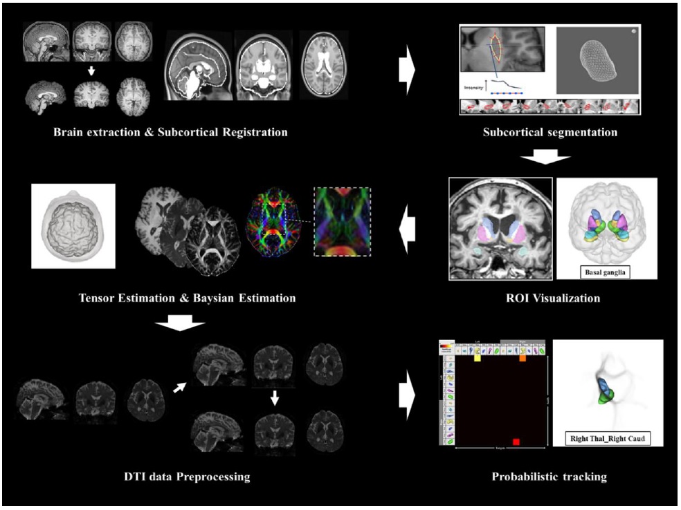
Depression in the elderly population has shown increased likelihood of neurological disorders due to structural changes in the subcortical area. However, further investigation into depression related subcortical changes is needed due to mismatches in structural analysis results between studies as well as scarcities in research regarding subcortical connectivity patterns of subclinical depression populations. This study aims to investigate structural differences in subcortical regions of aged participants with subclinical depression using 3Tesla MRI. In structural analysis, volumes of each subcortical region were measured to observe the volumetric difference and asymmetry between groups, but no significant difference was found. In addition, fractional anisotropy (FA) and apparent diffusion coefficient (ADC) did not show any significant differences between groups. Structural analysis using probabilistic tractography indicated that the connection strength between left nucleus accumbens-right hippocampus, and right thalamus-right caudate was higher in the control group than the subclinical depression group. The differences in subcortical connection strength of subclinical depression groups, have shown to correlate with emotional and cognitive disorders, such as anxiety and memory impairment. We believe that the analysis of structural differences and cross-regional network measures in subcortical structures can help identify neurophysiological changes occurring in subclinical depression.
Graphical Abstract
Keywords: Subclinical depression, Brain, Basal ganglia, Volumetric, Diffusion tensor imaging, Tractography