Articles

Article Tools
 View Full Text View Full Text |
 Abstract Abstract |
 Article as PDF Article as PDF |
 Print this Article Print this Article |
 Pubmed Pubmed |
 PMC PMC |
 PubReader PubReader |
 Export to Citation Export to Citation |
 Email Alerts Email Alerts |
 Open Access Open Access |
Share this article on :


Stats or Metrics
Article
Short Communication
Exp Neurobiol 2022; 31(2): 89-96
Published online April 30, 2022
https://doi.org/10.5607/en21019
© The Korean Society for Brain and Neural Sciences
Neonatal Mice Spinal Cord Interneurons Send Axons through the Dorsal Roots
Laura Paulina Osuna-Carrasco3, Sergio Horacio Dueñas-Jiménez1, Carmen Toro-Castillo3, Braniff De la Torre3, Irene Aguilar-García5, Jonatan Alpirez1, Luis Castillo2 and Judith Marcela Dueñas-Jiménez4*
1Department of Neuroscience, CUCS, University of Guadalajara, Guadalajara 44340, 2Basic Center, Autonomous University of Aguascalientes, Aguascalientes 20131, 3Department of Translational Bioengineering, CUCEI, University of Guadalajara, Guadalajara 44430, 4Department of Physiology, CUCS, University of Guadalajara, Guadalajara 44340, 5Department of Molecular and Genomics, CUCS, University of Guadalajara, Guadalajara 44340, México
Correspondence to: *To whom correspondence should be addressed.
TEL: 52-3310585313, FAX: 52-3310585200
e-mail: mduenas@cucs.udg.mx
This is an Open Access article distributed under the terms of the Creative Commons Attribution Non-Commercial License (http://creativecommons.org/licenses/by-nc/4.0) which permits unrestricted non-commercial use, distribution, and reproduction in any medium, provided the original work is properly cited.
Abstract
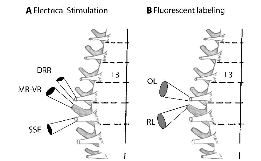
Spontaneous interneuron activity plays a critical role in developing neuronal networks. Discharges conducted antidromically along the dorsal root (DR) precede those from the ventral root’s (VR) motoneurons. This work studied whether spinal interneurons project axons into the neonate’s dorsal roots. Experiments were carried out in postnatal Swiss-Webster mice. We utilized a staining technique and found that interneurons in the spinal cord’s dorsal horn send axons through the dorsal roots. In vitro electrophysiological recordings showed antidromic action potentials (dorsal root reflex; DRR) produced by depolarizing the primary afferent terminals. These reflexes appeared by stimulating the adjacent dorsal roots. We found that bicuculline reduced the DRR evoked by L5 dorsal root stimulation when recording from the L4 dorsal root. Simultaneously, the monosynaptic reflex (MR) in the L5 ventral root was not affected; nevertheless, a long-lasting after-discharge appeared. The addition of 2-amino-5 phosphonovaleric acid (AP5), an NMDA receptor antagonist, abolished the MR without changing the after-discharge. The absence of DRR and MR facilitated single action potentials in the dorsal and ventral roots that persisted even in low Ca2+ concentrations. The results suggest that firing interneurons could send their axons through the dorsal roots. These interneurons could activate motoneurons producing individual spikes recorded in the ventral roots. Identifying these interneurons and the persistence of their neuronal connectivity in adulthood remains to be established.
Graphical Abstract
Keywords: Spinal cord interneurons, Antidromic activity, Dorsal root reflex, Monosynaptic reflex