Articles

Article Tools
 View Full Text View Full Text |
 Abstract Abstract |
 Article as PDF Article as PDF |
 Print this Article Print this Article |
 Pubmed Pubmed |
 PMC PMC |
 PubReader PubReader |
 Export to Citation Export to Citation |
 Email Alerts Email Alerts |
 Open Access Open Access |
Share this article on :


Stats or Metrics
Article
Review Article
Exp Neurobiol 2022; 31(3): 131-146
Published online June 30, 2022
https://doi.org/10.5607/en22015
© The Korean Society for Brain and Neural Sciences
Cranial and Spinal Window Preparation for in vivo Optical Neuroimaging in Rodents and Related Experimental Techniques
Chanmi Yeon1, Jeong Myo Im1, Minsung Kim1, Young Ro Kim2,3 and Euiheon Chung1,4,5*
1Department of Biomedical Science and Engineering, Gwangju Institute of Science and Technology, Gwangju 61005, Korea, 2Athinoula A. Martinos Center for Biomedical Imaging, Massachusetts General Hospital, Charlestown, MA 02129, 3Department of Radiology, Harvard Medical School, Boston, MA 02115, USA, 4AI Graduate School, Gwangju Institute of Science and Technology, Gwangju 61005, 5Research Center for Photon Science Technology, Gwangju Institute of Science and Technology, Gwangju 61005, Korea
Correspondence to: *To whom correspondence should be addressed.
TEL: 82-62-715-2753, FAX: 82-62-715-5309
e-mail: ogong50@gist.ac.kr
This is an Open Access article distributed under the terms of the Creative Commons Attribution Non-Commercial License (http://creativecommons.org/licenses/by-nc/4.0) which permits unrestricted non-commercial use, distribution, and reproduction in any medium, provided the original work is properly cited.
Abstract
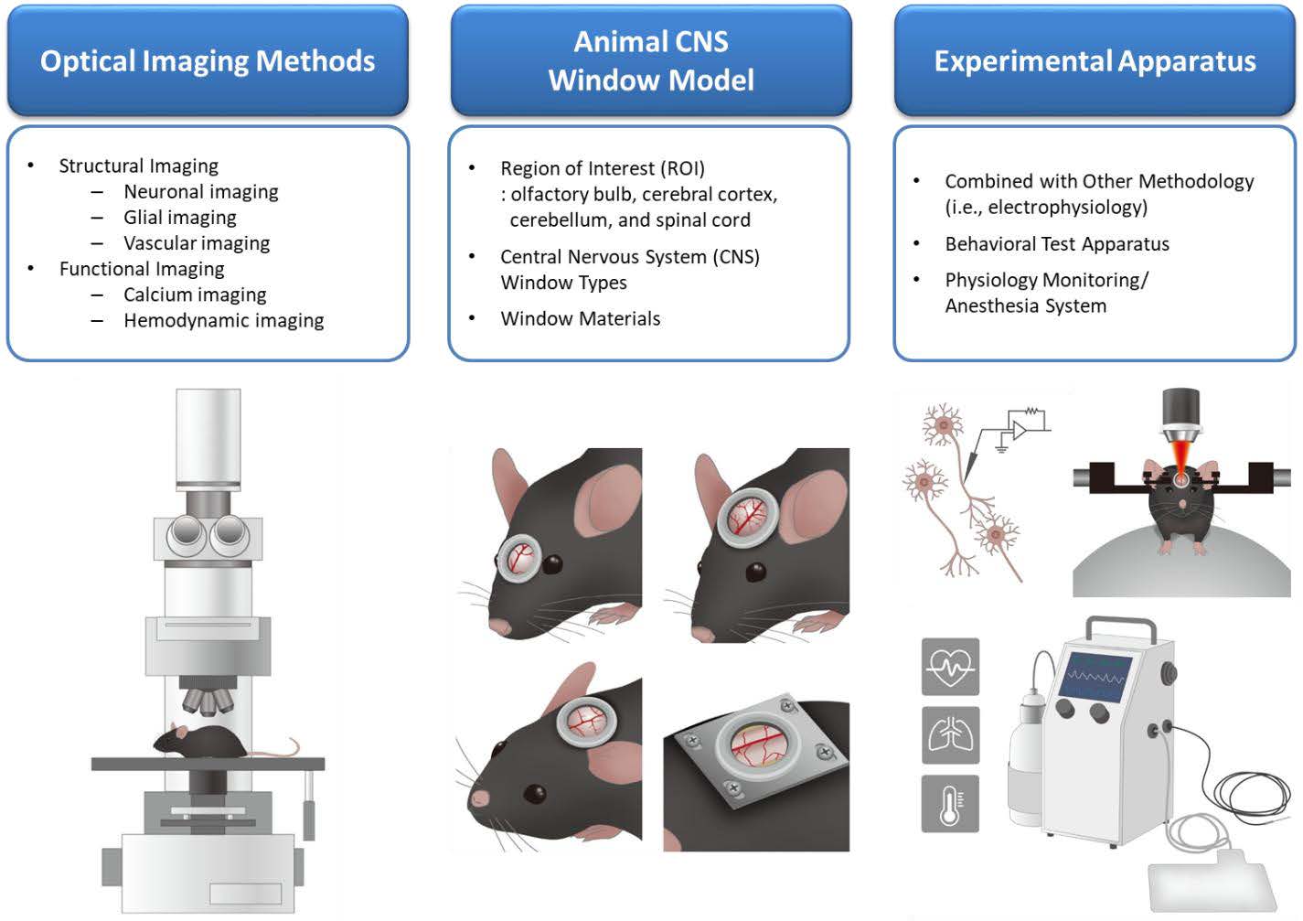
Optical neuroimaging provides an effective neuroscience tool for multi-scale investigation of the neural structures and functions, ranging from molecular, cellular activities to the inter-regional connectivity assessment. Amongst experimental preparations, the implementation of an artificial window to the central nervous system (CNS) is primarily required for optical visualization of the CNS and associated brain activities through the opaque skin and bone. Either thinning down or removing portions of the skull or spine is necessary for unobstructed long-term in vivo observations, for which types of the cranial and spinal window and applied materials vary depending on the study objectives. As diversely useful, a window can be designed to accommodate other experimental methods such as electrophysiology or optogenetics. Moreover, auxiliary apparatuses would allow the recording in synchrony with behavior of large-scale brain connectivity signals across the CNS, such as olfactory bulb, cerebral cortex, cerebellum, and spinal cord. Such advancements in the cranial and spinal window have resulted in a paradigm shift in neuroscience, enabling in vivo investigation of the brain function and dysfunction at the microscopic, cellular level. This Review addresses the types and classifications of windows used in optical neuroimaging while describing how to perform in vivo studies using rodent models in combination with other experimental modalities during behavioral tests. The cranial and spinal window has enabled longitudinal examination of evolving neural mechanisms via in situ visualization of the brain. We expect transformable and multi-functional cranial and spinal windows to become commonplace in neuroscience laboratories, further facilitating advances in optical neuroimaging systems.
Graphical Abstract
Keywords: Neuroimaging, Functional neuroimaging, Optical imaging, Central nervous system, Craniotomy, Laboratory animal models