Articles

Article Tools
 View Full Text View Full Text |
 Abstract Abstract |
 Article as PDF Article as PDF |
 Print this Article Print this Article |
 Pubmed Pubmed |
 PMC PMC |
 PubReader PubReader |
 Export to Citation Export to Citation |
 Email Alerts Email Alerts |
 Open Access Open Access |
Share this article on :


Stats or Metrics
Article
Short Communication
Exp Neurobiol 2022; 31(5): 353-360
Published online October 31, 2022
https://doi.org/10.5607/en22031
© The Korean Society for Brain and Neural Sciences
Reduced Sulcal Depth in Central Sulcus of Major Depressive Disorder
Seung-Joon Shin1, Aram Kim1, Kyu-Man Han2, Woo-Suk Tae3* and Byung-Joo Ham2*
1Department of Biomedical Sciences, Korea University College of Medicine,
2Department of Psychiatry, Korea University Anam Hospital, Korea University College of Medicine,
3Brain Convergence Research Center, Korea University College of Medicine, Seoul 02841, Korea
Correspondence to: *To whom correspondence should be addressed.
Woo-Suk Tae, TEL: 82-2-2286-1055, FAX: 82-0504-202-4629
e-mail: wstae@korea.ac.kr
Byung-Joo Ham, TEL: 82-2-9206-6843, FAX: 82-2-9206-6843
e-mail: hambj@korea.ac.kr
This is an Open Access article distributed under the terms of the Creative Commons Attribution Non-Commercial License (http://creativecommons.org/licenses/by-nc/4.0) which permits unrestricted non-commercial use, distribution, and reproduction in any medium, provided the original work is properly cited.
Abstract
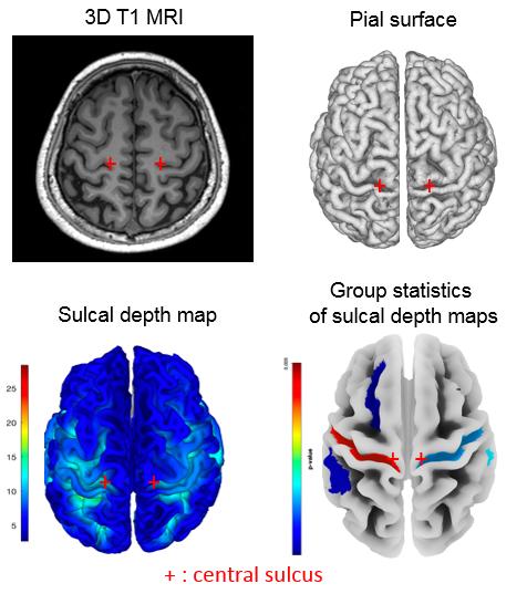
Major depressive disorder (MDD) is one of the most common psychiatric disorders, and present various symptoms such as the dysregulation of mood, cognition, and behavior. The purpose of the present study was to investigate the morphometric change in MDD patients by voxel-based morphometry (VBM) and sulcal depth analyses. Forty-six MDD patients (mean age, SD; 36.07±14.34), and 23 age- and sex-matched normal controls (NML) (mean age, SD; 36.78±14.42) were included. Coronal 3D T1 magnetic resonance imaging (MRI) was obtained with the resolution of isotropic 1.0 mm. To check morphological changes of brain, T1 MRIs were objectively processed by VBM and sulcal depth methods. In sulcal depth analysis, depressed patients showed reduced sulcal depth in the areas of left posterior ramus of the lateral sulcus, superior frontal sulcus, supramarginal gyrus, central sulcus (Rolando's fissure), and Heschl's gyrus. And right posterior ramus of the lateral sulcus, temporal plane of the superior temporal gyrus, anterior transverse collateral sulcus, and central sulcus (Rolando’s fissure) were also reduced compared to NML. But, VBM analyses did not showed significant finding. Reduced sulcal depth in the motor and emotion related areas were found in patients with MDD. Especially reduced sulcal depth in bilateral central sulci which are connecting between primary motor cortex and primary sensory cortex seems to be related with social and physical anhedonia in MDD.
Graphical Abstract
Keywords: Major depressive disorder, Magnetic resonance imaging, Voxel-based morphometry, Sulcal depth, Central sulcus