Articles

Article Tools
 View Full Text View Full Text |
 Abstract Abstract |
 Article as PDF Article as PDF |
 Print this Article Print this Article |
 Pubmed Pubmed |
 PMC PMC |
 PubReader PubReader |
 Export to Citation Export to Citation |
 Email Alerts Email Alerts |
 Open Access Open Access |
Share this article on :


Stats or Metrics
Article
Original Article
Exp Neurobiol 2022; 31(5): 299-306
Published online October 31, 2022
https://doi.org/10.5607/en22020
© The Korean Society for Brain and Neural Sciences
Synaptic Remodeling of the Auditory Cortex Following Bilateral Blindness: Evidence of Cross-modal Plasticity
Jae Joon Han1†, Tae-Soo Noh2†, Myung-Whan Suh2, Seung Ha Kim3,4, Doo Hee Kim2, Sang Jeong Kim3,4,5* and Seung Ha Oh2*
1Department of Otorhinolaryngology–Head and Neck Surgery, Soonchunhyang University College of Medicine, Seoul Hospital, Seoul 04401, 2Department of Otorhinolaryngology-Head and Neck Surgery, Seoul National University Hospital, Seoul National University College of Medicine, Seoul 03080, 3Department of Physiology, Seoul National University College of Medicine, Seoul 03080, 4Department of Biomedical Sciences, Seoul National University College of Medicine, Seoul 03080, 5Neuroscience Research Institute, Seoul National University College of Natural Sciences, Seoul 03080, Korea
Correspondence to: *To whom correspondence should be addressed.
Sang Jeong Kim, TEL: 82-2-740-8229, FAX: 82-2-763-9667
e-mail: sangjkim@snu.ac.kr
Seung Ha Oh, TEL: 82-2-2072-2442, FAX: 82-2-745-2387
e-mail: shaoh@snu.ac.kr
†These authors contributed equally to this article.
This is an Open Access article distributed under the terms of the Creative Commons Attribution Non-Commercial License (http://creativecommons.org/licenses/by-nc/4.0) which permits unrestricted non-commercial use, distribution, and reproduction in any medium, provided the original work is properly cited.
Abstract
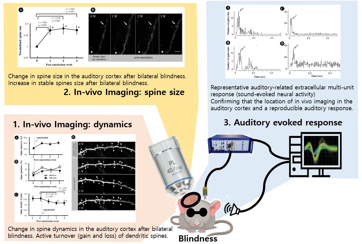
We aimed to evaluate structural dynamic changes of neurons in the auditory cortex after visual deprivation. We longitudinally tracked dendritic spines for 3 weeks after visual deprivation in vivo using a two-photon microscope. GFP-labeled dendritic spines in the auditory cortex were serially followed after bilateral enucleation. The turnover rate, density, and size of the spines in the dendrites were evaluated 1, 2, and 3 weeks after visual deprivation. The turnover rate of the dendritic spines in the auditory cortex increased at 1 week (20.1±7.3%) after bilateral enucleation compared to baseline (12.5±7.9%); the increase persisted for up to 3 weeks (20.9±11.0%). The spine loss rate was slightly higher than the spine gain rate. The average spine density (number of spines per 1 μm of dendrite) was significantly lower at 2 weeks (2W; 0.22±0.06 1/μm) and 3 W (0.22±0.08 1/μm) post-nucleation compared to baseline (0.026±0.09 1/μm). We evaluated the change of synaptic strength in the stable spines at each time point. The normalized spine size in the auditory cortex was significantly increased after bilateral blindness at 1 W postoperatively (1.36±0.92), 2 W postoperatively (1.40±1.18), and 3 W postoperatively (1.36±0.88) compared to baseline. Sensory deprivation resulted in remodeling of the neural circuitry in the spared cortex, via cross-modal plasticity in the direction of partial breakdown of synapses, and enhanced strength of the remaining synapses.
Graphical Abstract
Keywords: Auditory cortex, Cross-modal plasticity, Two-photon, Sensory deprivation