Articles

Article Tools
 View Full Text View Full Text |
 Abstract Abstract |
 Article as PDF Article as PDF |
 Print this Article Print this Article |
 Pubmed Pubmed |
 PMC PMC |
 PubReader PubReader |
 Export to Citation Export to Citation |
 Email Alerts Email Alerts |
 Open Access Open Access |
Supplementary
Share this article on :


Stats or Metrics
Article
Original Article
Exp Neurobiol 2023; 32(3): 181-194
Published online June 30, 2023
https://doi.org/10.5607/en23001
© The Korean Society for Brain and Neural Sciences
An Automated Cell Detection Method for TH-positive Dopaminergic Neurons in a Mouse Model of Parkinson’s Disease Using Convolutional Neural Networks
Doyun Kim1†, Myeong Seong Bak2,3†, Haney Park2,3, In Seon Baek2, Geehoon Chung1, Jae Hyun Park4, Sora Ahn5, Seon-Young Park1, Hyunsu Bae1, Hi-Joon Park5 and Sun Kwang Kim1,2,4*
1Department of Physiology, College of Korean Medicine, Kyung Hee University, Seoul 02447, 2Department of Science in Korean Medicine, Graduate School, Kyung Hee University, Seoul 02447, 3Department of AI and Data Analysis, Neurogrin Inc., Seoul 02455, 4Department of East-West Medicine, Graduate School, Kyung Hee University, Seoul 02447, 5Acupuncture & Meridian Science Research Center, Kyung Hee University, Seoul 02447, Korea
Correspondence to: *To whom correspondence should be addressed.
TEL: 82-2-961-0323, FAX: 82-2-961-0333
e-mail: skkim77@khu.ac.kr
†These authors contributed equally to this article.
This is an Open Access article distributed under the terms of the Creative Commons Attribution Non-Commercial License (http://creativecommons.org/licenses/by-nc/4.0) which permits unrestricted non-commercial use, distribution, and reproduction in any medium, provided the original work is properly cited.
Abstract
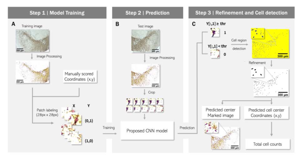
Quantification of tyrosine hydroxylase (TH)-positive neurons is essential for the preclinical study of Parkinson’s disease (PD). However, manual analysis of immunohistochemical (IHC) images is labor-intensive and has less reproducibility due to the lack of objectivity. Therefore, several automated methods of IHC image analysis have been proposed, although they have limitations of low accuracy and difficulties in practical use. Here, we developed a convolutional neural network-based machine learning algorithm for TH+ cell counting. The developed analytical tool showed higher accuracy than the conventional methods and could be used under diverse experimental conditions of image staining intensity, brightness, and contrast. Our automated cell detection algorithm is available for free and has an intelligible graphical user interface for cell counting to assist practical applications. Overall, we expect that the proposed TH+ cell counting tool will promote preclinical PD research by saving time and enabling objective analysis of IHC images.
Graphical Abstract
Keywords: Parkinson’s disease, Mice, Dopaminergic neurons, Deep learning, Neural networks, Cell count